Bromine in PDB 8hu5: Crystal Structure of Dna Octamer Containing Guna[Me,Tbu]
Protein crystallography data
The structure of Crystal Structure of Dna Octamer Containing Guna[Me,Tbu], PDB code: 8hu5
was solved by
H.Aoyama,
H.Obika,
T.Yamaguchi,
with X-Ray Crystallography technique. A brief refinement statistics is given in the table below:
| Resolution Low / High (Å) | 21.10 / 0.93 |
| Space group | P 43 21 2 |
| Cell size a, b, c (Å), α, β, γ (°) | 42.494, 42.494, 24.315, 90, 90, 90 |
| R / Rfree (%) | 19.2 / 21.3 |
Bromine Binding Sites:
The binding sites of Bromine atom in the Crystal Structure of Dna Octamer Containing Guna[Me,Tbu]
(pdb code 8hu5). This binding sites where shown within
5.0 Angstroms radius around Bromine atom.
In total only one binding site of Bromine was determined in the Crystal Structure of Dna Octamer Containing Guna[Me,Tbu], PDB code: 8hu5:
In total only one binding site of Bromine was determined in the Crystal Structure of Dna Octamer Containing Guna[Me,Tbu], PDB code: 8hu5:
Bromine binding site 1 out of 1 in 8hu5
Go back to
Bromine binding site 1 out
of 1 in the Crystal Structure of Dna Octamer Containing Guna[Me,Tbu]
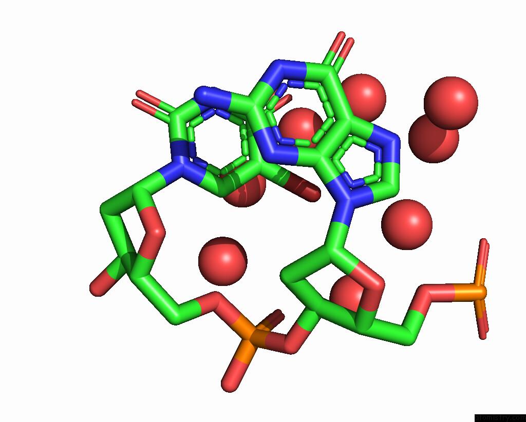
Mono view
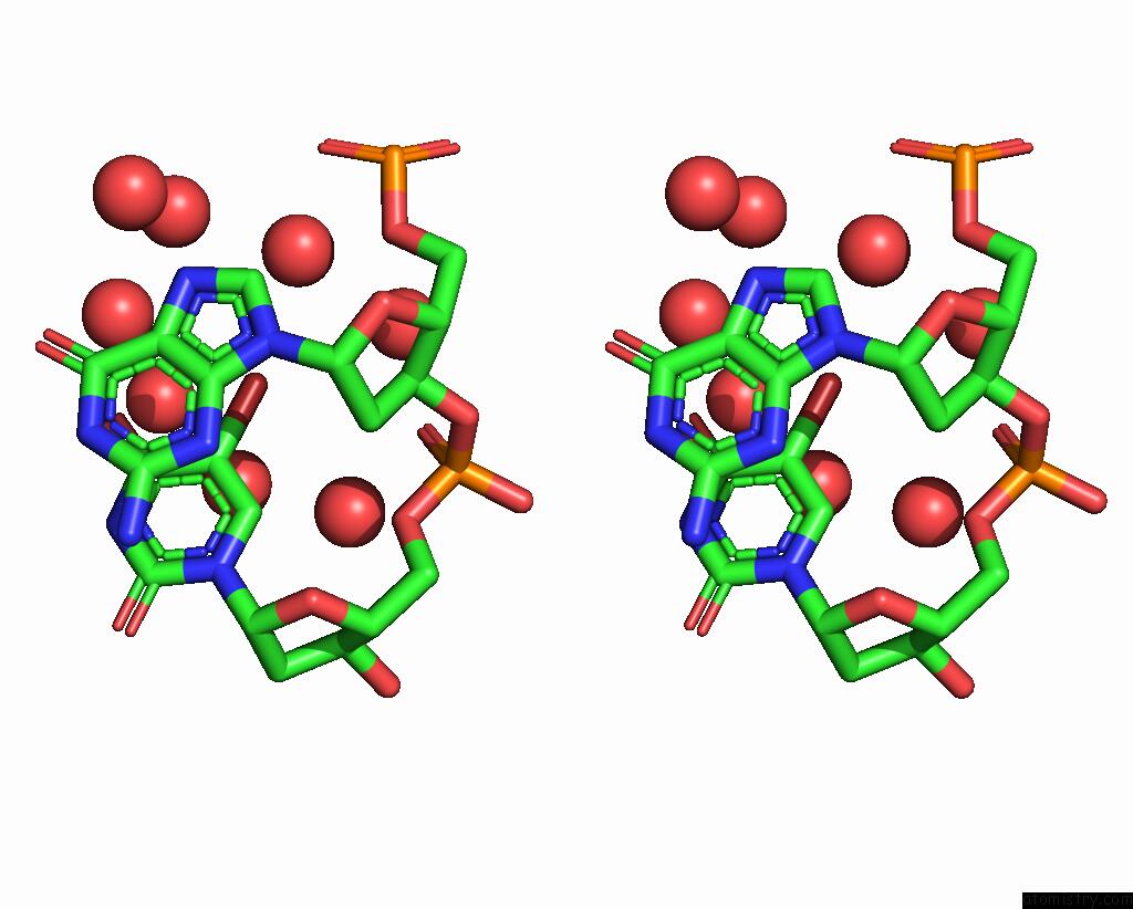
Stereo pair view
Mono view
Stereo pair view
A full contact list of Bromine with other atoms in the Br binding
site number 1 of Crystal Structure of Dna Octamer Containing Guna[Me,Tbu] within 5.0Å range:
|
Reference:
T.Yamaguchi,
N.Horie,
H.Aoyama,
S.Kumagai,
S.Obika.
Mechanism of the Extremely High Duplex-Forming Ability of Oligonucleotides Modified with N-Tert-Butylguanidine- or N-Tert-Butyl-N'-Methylguanidine-Bridged Nucleic Acids. Nucleic Acids Res. 2023.
ISSN: ESSN 1362-4962
PubMed: 37462081
DOI: 10.1093/NAR/GKAD608
Page generated: Thu Jul 11 05:16:54 2024
ISSN: ESSN 1362-4962
PubMed: 37462081
DOI: 10.1093/NAR/GKAD608
Last articles
Zn in 9MJ5Zn in 9HNW
Zn in 9G0L
Zn in 9FNE
Zn in 9DZN
Zn in 9E0I
Zn in 9D32
Zn in 9DAK
Zn in 8ZXC
Zn in 8ZUF