Bromine »
PDB 1e7b-1ih6 »
1hd2 »
Bromine in PDB 1hd2: Human Peroxiredoxin 5
Protein crystallography data
The structure of Human Peroxiredoxin 5, PDB code: 1hd2
was solved by
J.P.Declercq,
C.Evrard,
with X-Ray Crystallography technique. A brief refinement statistics is given in the table below:
| Resolution Low / High (Å) | 16.00 / 1.50 |
| Space group | P 41 21 2 |
| Cell size a, b, c (Å), α, β, γ (°) | 66.607, 66.607, 123.327, 90.00, 90.00, 90.00 |
| R / Rfree (%) | 13.3 / 16.5 |
Bromine Binding Sites:
The binding sites of Bromine atom in the Human Peroxiredoxin 5
(pdb code 1hd2). This binding sites where shown within
5.0 Angstroms radius around Bromine atom.
In total 5 binding sites of Bromine where determined in the Human Peroxiredoxin 5, PDB code: 1hd2:
Jump to Bromine binding site number: 1; 2; 3; 4; 5;
In total 5 binding sites of Bromine where determined in the Human Peroxiredoxin 5, PDB code: 1hd2:
Jump to Bromine binding site number: 1; 2; 3; 4; 5;
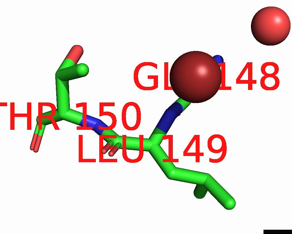
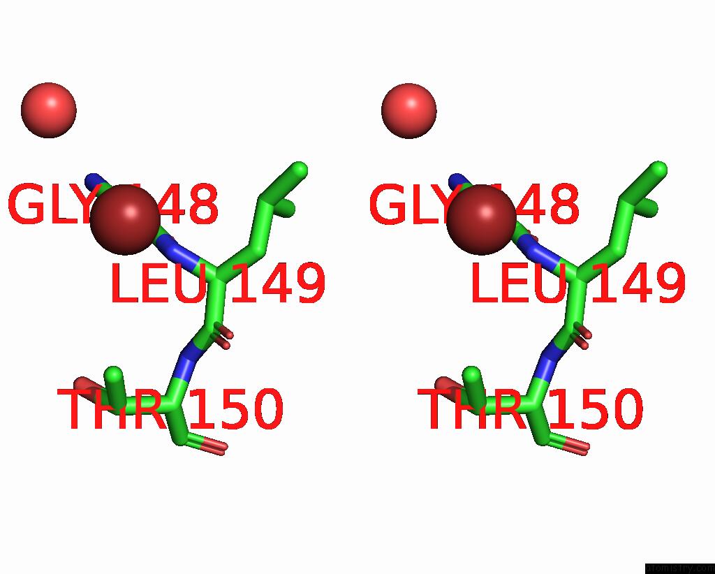
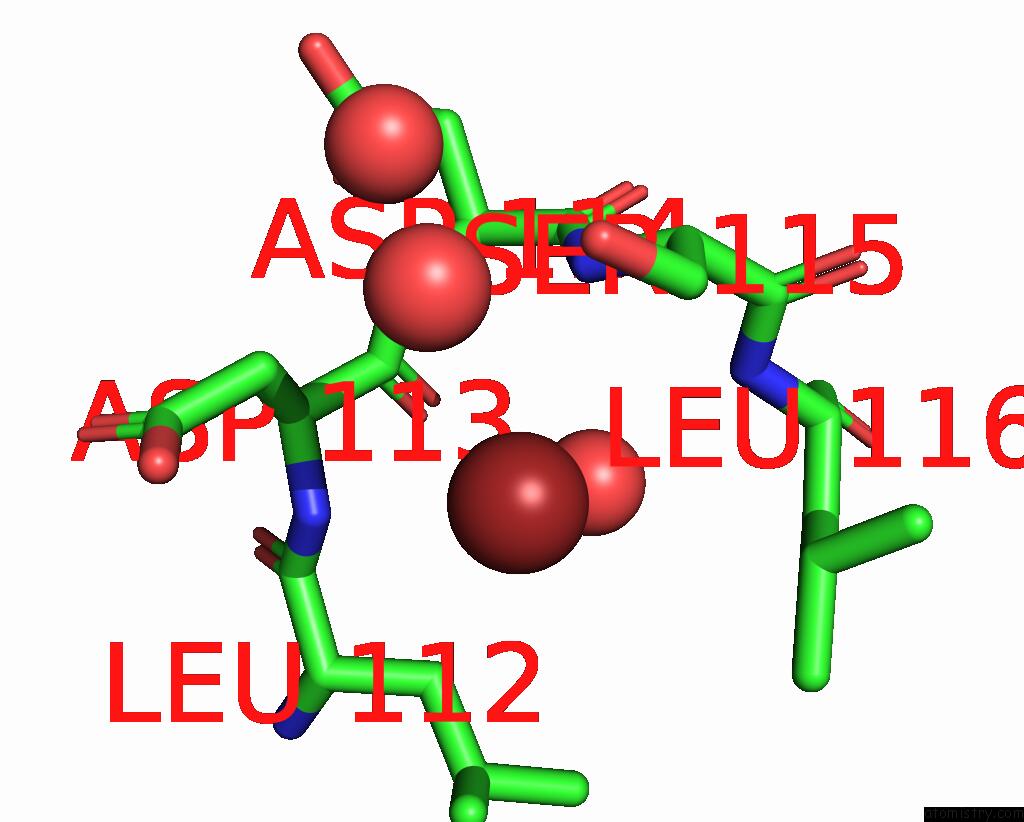
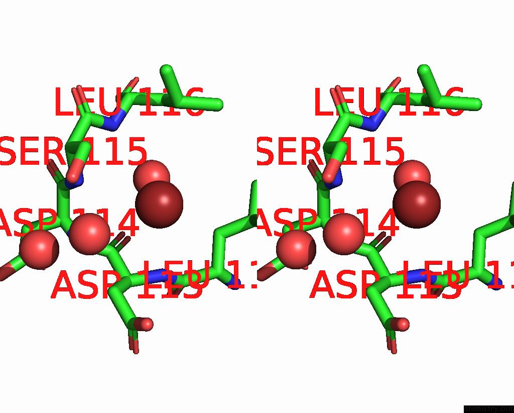
Bromine binding site 1 out of 5 in 1hd2
Go back to
Bromine binding site 1 out
of 5 in the Human Peroxiredoxin 5
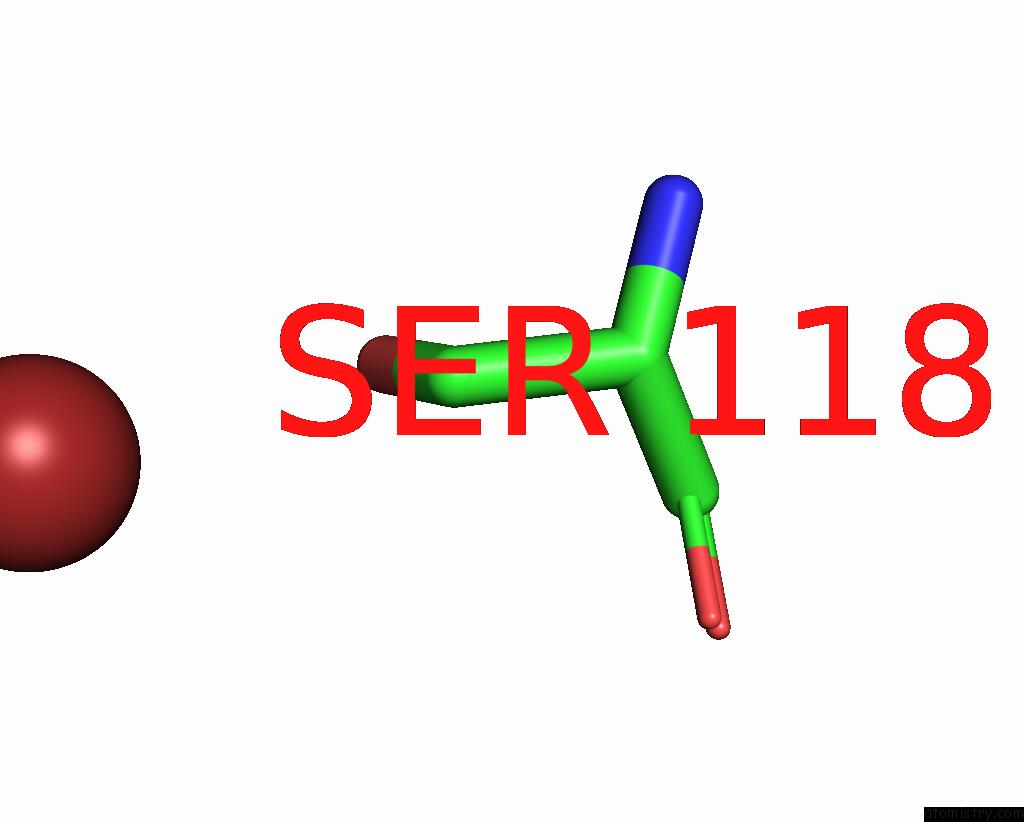
Mono view
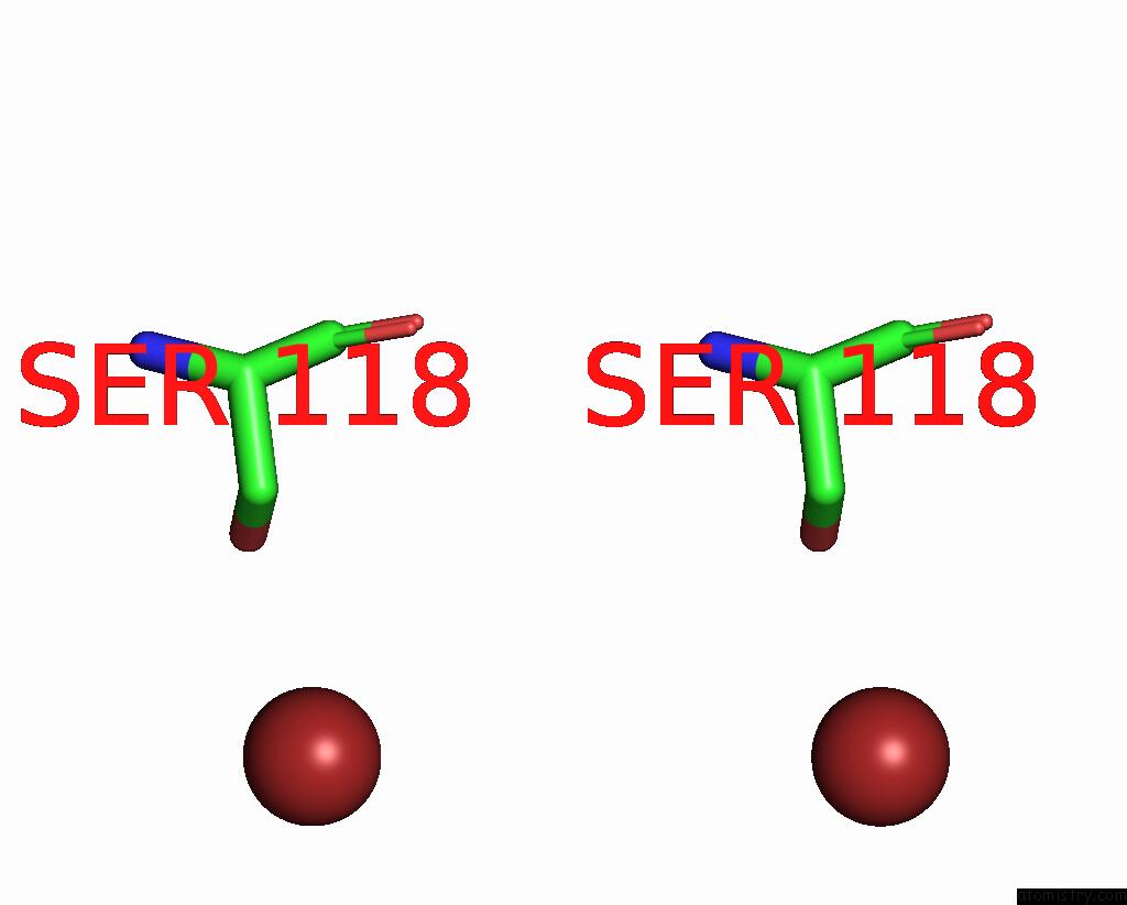
Stereo pair view
Mono view
Stereo pair view
A full contact list of Bromine with other atoms in the Br binding
site number 1 of Human Peroxiredoxin 5 within 5.0Å range:
|
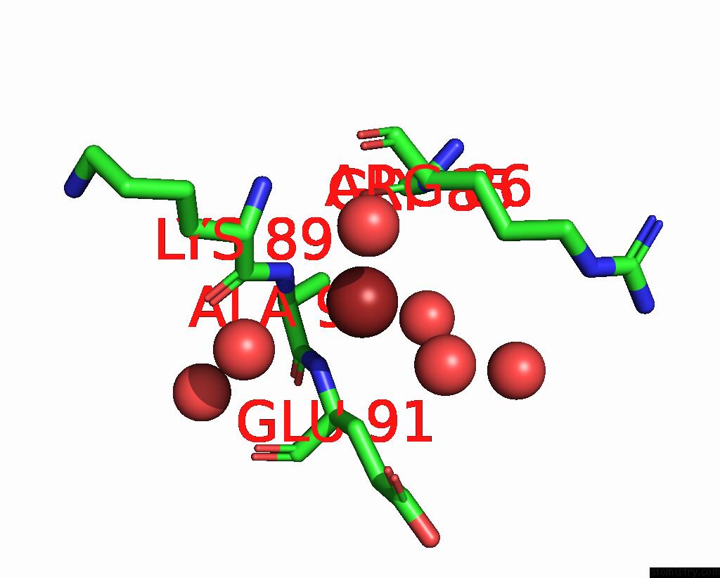
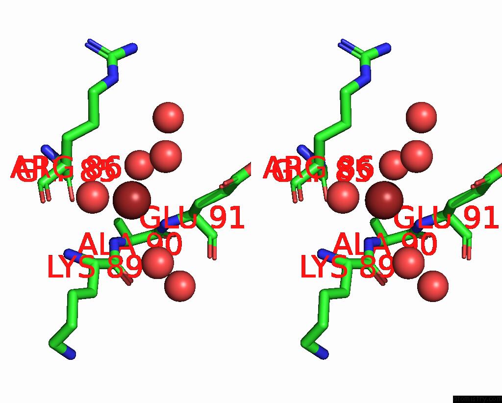
Bromine binding site 2 out of 5 in 1hd2
Go back to
Bromine binding site 2 out
of 5 in the Human Peroxiredoxin 5
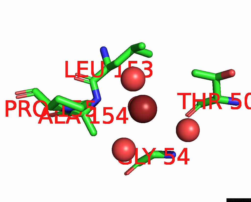
Mono view
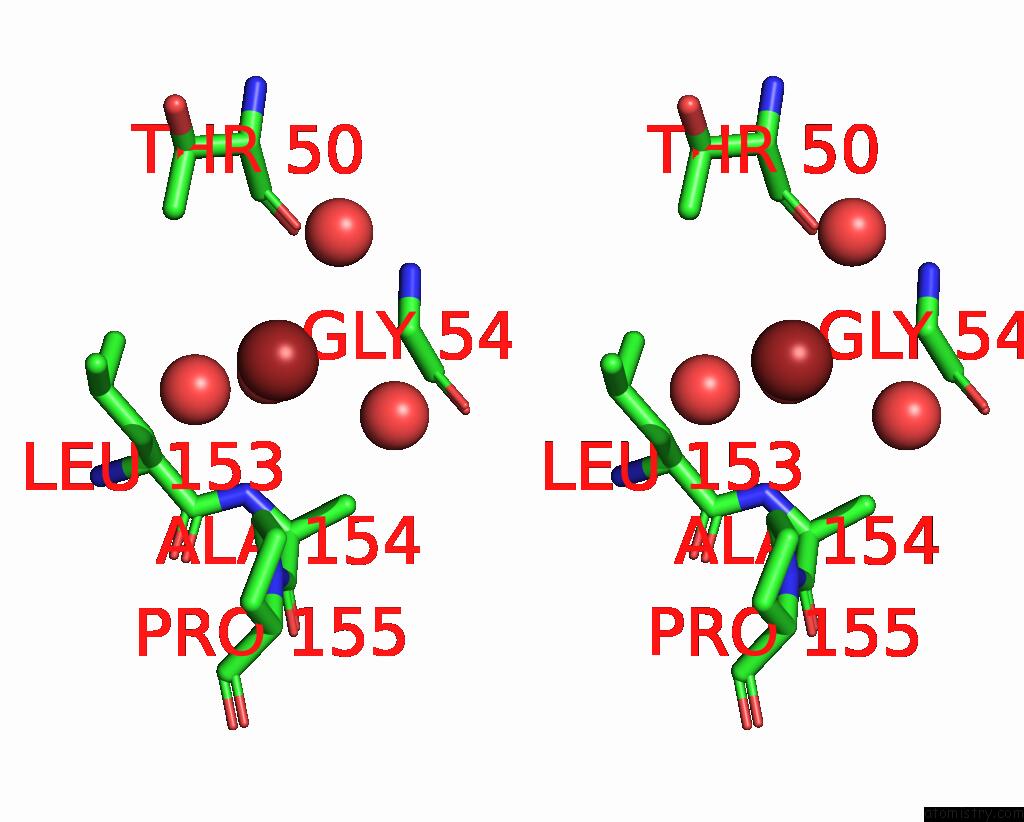
Stereo pair view
Mono view
Stereo pair view
A full contact list of Bromine with other atoms in the Br binding
site number 2 of Human Peroxiredoxin 5 within 5.0Å range:
|
Bromine binding site 3 out of 5 in 1hd2
Go back to
Bromine binding site 3 out
of 5 in the Human Peroxiredoxin 5
Mono view
Stereo pair view
Mono view
Stereo pair view
A full contact list of Bromine with other atoms in the Br binding
site number 3 of Human Peroxiredoxin 5 within 5.0Å range:
|
Bromine binding site 4 out of 5 in 1hd2
Go back to
Bromine binding site 4 out
of 5 in the Human Peroxiredoxin 5
Mono view
Stereo pair view
Mono view
Stereo pair view
A full contact list of Bromine with other atoms in the Br binding
site number 4 of Human Peroxiredoxin 5 within 5.0Å range:
|
Bromine binding site 5 out of 5 in 1hd2
Go back to
Bromine binding site 5 out
of 5 in the Human Peroxiredoxin 5
Mono view
Stereo pair view
Mono view
Stereo pair view
A full contact list of Bromine with other atoms in the Br binding
site number 5 of Human Peroxiredoxin 5 within 5.0Å range:
|
Reference:
J.P.Declercq,
C.Evrard,
A.Clippe,
D.V.Stricht,
A.Bernard,
B.Knoops.
Crystal Structure of Human Peroxiredoxin 5, A Novel Type of Mammalian Peroxiredoxin at 1.5 A Resolution. J.Mol.Biol. V. 311 751 2001.
ISSN: ISSN 0022-2836
PubMed: 11518528
DOI: 10.1006/JMBI.2001.4853
Page generated: Mon Jul 7 03:17:08 2025
ISSN: ISSN 0022-2836
PubMed: 11518528
DOI: 10.1006/JMBI.2001.4853
Last articles
F in 7G0TF in 7FY8
F in 7FZH
F in 7G03
F in 7FZ4
F in 7FYY
F in 7FZ2
F in 7FXG
F in 7FYQ
F in 7FXS