Bromine »
PDB 1iha-1m9r »
1m5d »
Bromine in PDB 1m5d: X-Ray Structure of the GLUR2 Ligand Binding Core (S1S2J-Y702F) in Complex with Br-Hibo at 1.73 A Resolution
Protein crystallography data
The structure of X-Ray Structure of the GLUR2 Ligand Binding Core (S1S2J-Y702F) in Complex with Br-Hibo at 1.73 A Resolution, PDB code: 1m5d
was solved by
A.Hogner,
J.S.Kastrup,
R.Jin,
T.Liljefors,
M.L.Mayer,
J.Egebjerg,
I.K.Larsen,
E.Gouaux,
with X-Ray Crystallography technique. A brief refinement statistics is given in the table below:
| Resolution Low / High (Å) | 19.59 / 1.73 |
| Space group | P 21 21 2 |
| Cell size a, b, c (Å), α, β, γ (°) | 87.139, 63.681, 47.914, 90.00, 90.00, 90.00 |
| R / Rfree (%) | 18.6 / 21.5 |
Bromine Binding Sites:
The binding sites of Bromine atom in the X-Ray Structure of the GLUR2 Ligand Binding Core (S1S2J-Y702F) in Complex with Br-Hibo at 1.73 A Resolution
(pdb code 1m5d). This binding sites where shown within
5.0 Angstroms radius around Bromine atom.
In total only one binding site of Bromine was determined in the X-Ray Structure of the GLUR2 Ligand Binding Core (S1S2J-Y702F) in Complex with Br-Hibo at 1.73 A Resolution, PDB code: 1m5d:
In total only one binding site of Bromine was determined in the X-Ray Structure of the GLUR2 Ligand Binding Core (S1S2J-Y702F) in Complex with Br-Hibo at 1.73 A Resolution, PDB code: 1m5d:
Bromine binding site 1 out of 1 in 1m5d
Go back to
Bromine binding site 1 out
of 1 in the X-Ray Structure of the GLUR2 Ligand Binding Core (S1S2J-Y702F) in Complex with Br-Hibo at 1.73 A Resolution
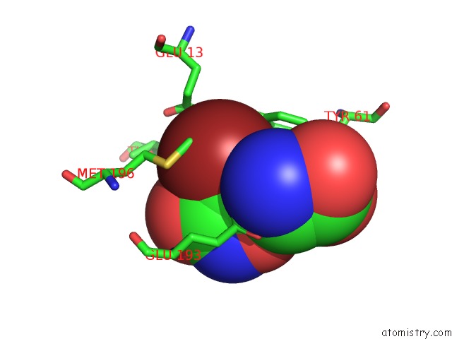
Mono view
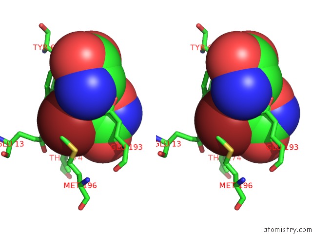
Stereo pair view
Mono view
Stereo pair view
A full contact list of Bromine with other atoms in the Br binding
site number 1 of X-Ray Structure of the GLUR2 Ligand Binding Core (S1S2J-Y702F) in Complex with Br-Hibo at 1.73 A Resolution within 5.0Å range:
|
Reference:
A.Hogner,
J.S.Kastrup,
R.Jin,
T.Liljefors,
M.L.Mayer,
J.Egebjerg,
I.K.Larsen,
E.Gouaux.
Structural Basis For Ampa Receptor Activation and Ligand Selectivity: Crystal Structures of Five Agonist Complexes with the GLUR2 Ligand-Binding Core J.Mol.Biol. V. 322 93 2002.
ISSN: ISSN 0022-2836
PubMed: 12215417
DOI: 10.1016/S0022-2836(02)00650-2
Page generated: Mon Jul 7 03:25:57 2025
ISSN: ISSN 0022-2836
PubMed: 12215417
DOI: 10.1016/S0022-2836(02)00650-2
Last articles
Br in 5O0BBr in 5O0J
Br in 5NZD
Br in 5NZ3
Br in 5MZT
Br in 5NZ6
Br in 5NY8
Br in 5NXI
Br in 5NWQ
Br in 5NRA