Bromine »
PDB 1m9t-1p1y »
1mqh »
Bromine in PDB 1mqh: Crystal Structure of the GLUR2 Ligand Binding Core (S1S2J) in Complex with Bromo-Willardiine at 1.8 Angstroms Resolution
Protein crystallography data
The structure of Crystal Structure of the GLUR2 Ligand Binding Core (S1S2J) in Complex with Bromo-Willardiine at 1.8 Angstroms Resolution, PDB code: 1mqh
was solved by
R.Jin,
T.G.Banke,
M.L.Mayer,
S.F.Traynelis,
E.Gouaux,
with X-Ray Crystallography technique. A brief refinement statistics is given in the table below:
| Resolution Low / High (Å) | 27.60 / 1.80 |
| Space group | P 21 21 2 |
| Cell size a, b, c (Å), α, β, γ (°) | 63.786, 91.851, 48.377, 90.00, 90.00, 90.00 |
| R / Rfree (%) | 19.8 / 23.1 |
Bromine Binding Sites:
The binding sites of Bromine atom in the Crystal Structure of the GLUR2 Ligand Binding Core (S1S2J) in Complex with Bromo-Willardiine at 1.8 Angstroms Resolution
(pdb code 1mqh). This binding sites where shown within
5.0 Angstroms radius around Bromine atom.
In total only one binding site of Bromine was determined in the Crystal Structure of the GLUR2 Ligand Binding Core (S1S2J) in Complex with Bromo-Willardiine at 1.8 Angstroms Resolution, PDB code: 1mqh:
In total only one binding site of Bromine was determined in the Crystal Structure of the GLUR2 Ligand Binding Core (S1S2J) in Complex with Bromo-Willardiine at 1.8 Angstroms Resolution, PDB code: 1mqh:
Bromine binding site 1 out of 1 in 1mqh
Go back to
Bromine binding site 1 out
of 1 in the Crystal Structure of the GLUR2 Ligand Binding Core (S1S2J) in Complex with Bromo-Willardiine at 1.8 Angstroms Resolution
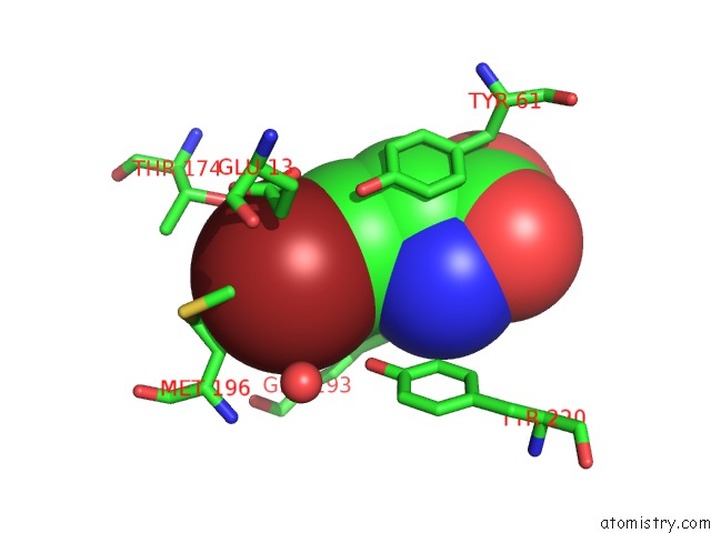
Mono view
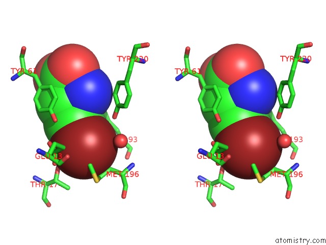
Stereo pair view
Mono view
Stereo pair view
A full contact list of Bromine with other atoms in the Br binding
site number 1 of Crystal Structure of the GLUR2 Ligand Binding Core (S1S2J) in Complex with Bromo-Willardiine at 1.8 Angstroms Resolution within 5.0Å range:
|
Reference:
R.Jin,
T.G.Banke,
M.L.Mayer,
S.F.Traynelis,
E.Gouaux.
Structural Basis For Partial Agonist Action at Ionotropic Glutamate Receptors Nat.Neurosci. V. 6 803 2003.
ISSN: ISSN 1097-6256
PubMed: 12872125
DOI: 10.1038/NN1091
Page generated: Mon Jul 7 03:30:06 2025
ISSN: ISSN 1097-6256
PubMed: 12872125
DOI: 10.1038/NN1091
Last articles
Br in 6JCJBr in 6JKN
Br in 6JJE
Br in 6I6T
Br in 6IYQ
Br in 6I3A
Br in 6IC2
Br in 6IBV
Br in 6I6G
Br in 6HTM