Bromine »
PDB 1p2x-1to3 »
1rer »
Bromine in PDB 1rer: Crystal Structure of the Homotrimer of Fusion Glycoprotein E1 From Semliki Forest Virus.
Protein crystallography data
The structure of Crystal Structure of the Homotrimer of Fusion Glycoprotein E1 From Semliki Forest Virus., PDB code: 1rer
was solved by
D.L.Gibbons,
M.C.Vaney,
A.Roussel,
A.Vigouroux,
B.Reilly,
M.Kielian,
F.A.Rey,
with X-Ray Crystallography technique. A brief refinement statistics is given in the table below:
| Resolution Low / High (Å) | 20.00 / 3.20 |
| Space group | P 31 2 1 |
| Cell size a, b, c (Å), α, β, γ (°) | 198.197, 198.197, 116.250, 90.00, 90.00, 120.00 |
| R / Rfree (%) | 26.5 / 28.5 |
Other elements in 1rer:
The structure of Crystal Structure of the Homotrimer of Fusion Glycoprotein E1 From Semliki Forest Virus. also contains other interesting chemical elements:
| Holmium | (Ho) | 4 atoms |
Bromine Binding Sites:
The binding sites of Bromine atom in the Crystal Structure of the Homotrimer of Fusion Glycoprotein E1 From Semliki Forest Virus.
(pdb code 1rer). This binding sites where shown within
5.0 Angstroms radius around Bromine atom.
In total 3 binding sites of Bromine where determined in the Crystal Structure of the Homotrimer of Fusion Glycoprotein E1 From Semliki Forest Virus., PDB code: 1rer:
Jump to Bromine binding site number: 1; 2; 3;
In total 3 binding sites of Bromine where determined in the Crystal Structure of the Homotrimer of Fusion Glycoprotein E1 From Semliki Forest Virus., PDB code: 1rer:
Jump to Bromine binding site number: 1; 2; 3;
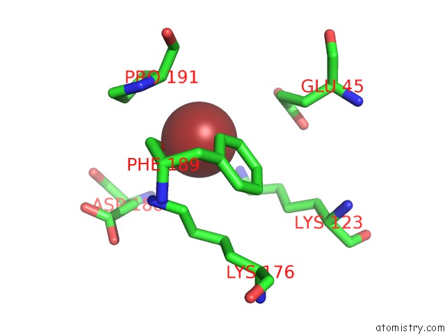
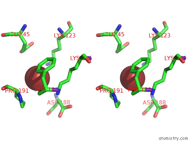
Bromine binding site 1 out of 3 in 1rer
Go back to
Bromine binding site 1 out
of 3 in the Crystal Structure of the Homotrimer of Fusion Glycoprotein E1 From Semliki Forest Virus.
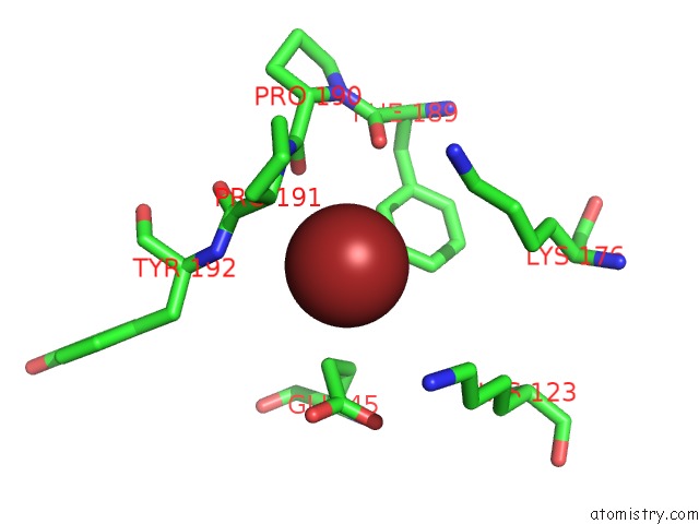
Mono view
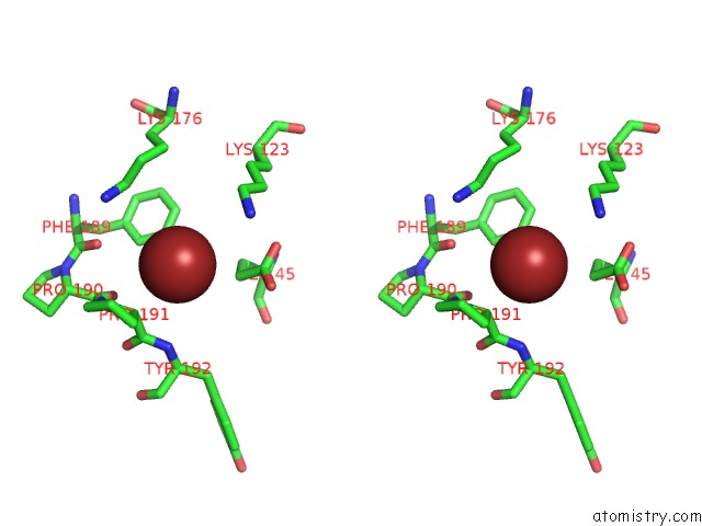
Stereo pair view
Mono view
Stereo pair view
A full contact list of Bromine with other atoms in the Br binding
site number 1 of Crystal Structure of the Homotrimer of Fusion Glycoprotein E1 From Semliki Forest Virus. within 5.0Å range:
|
Bromine binding site 2 out of 3 in 1rer
Go back to
Bromine binding site 2 out
of 3 in the Crystal Structure of the Homotrimer of Fusion Glycoprotein E1 From Semliki Forest Virus.
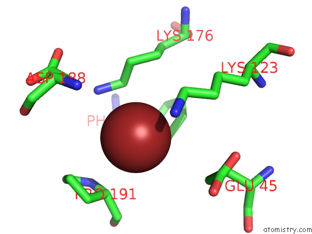
Mono view
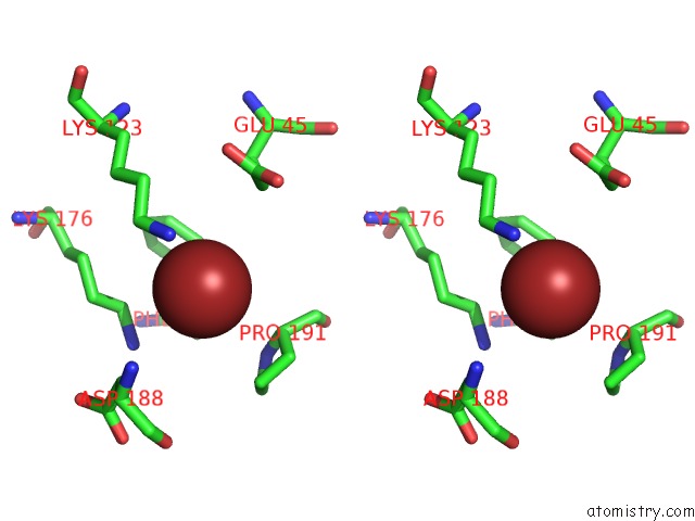
Stereo pair view
Mono view
Stereo pair view
A full contact list of Bromine with other atoms in the Br binding
site number 2 of Crystal Structure of the Homotrimer of Fusion Glycoprotein E1 From Semliki Forest Virus. within 5.0Å range:
|
Bromine binding site 3 out of 3 in 1rer
Go back to
Bromine binding site 3 out
of 3 in the Crystal Structure of the Homotrimer of Fusion Glycoprotein E1 From Semliki Forest Virus.
Mono view
Stereo pair view
Mono view
Stereo pair view
A full contact list of Bromine with other atoms in the Br binding
site number 3 of Crystal Structure of the Homotrimer of Fusion Glycoprotein E1 From Semliki Forest Virus. within 5.0Å range:
|
Reference:
D.L.Gibbons,
M.C.Vaney,
A.Roussel,
A.Vigouroux,
B.Reilly,
J.Lepault,
M.Kielian,
F.A.Rey.
Conformational Change and Protein-Protein Interactions of the Fusion Protein of Semliki Forest Virus. Nature V. 427 320 2004.
ISSN: ISSN 0028-0836
PubMed: 14737160
DOI: 10.1038/NATURE02239
Page generated: Wed Jul 10 17:16:36 2024
ISSN: ISSN 0028-0836
PubMed: 14737160
DOI: 10.1038/NATURE02239
Last articles
Zn in 9J0NZn in 9J0O
Zn in 9J0P
Zn in 9FJX
Zn in 9EKB
Zn in 9C0F
Zn in 9CAH
Zn in 9CH0
Zn in 9CH3
Zn in 9CH1