Bromine »
PDB 2civ-2hb1 »
2h34 »
Bromine in PDB 2h34: Apoenzyme Crystal Structure of the Tuberculosis Serine/Threonine Kinase, Pkne
Enzymatic activity of Apoenzyme Crystal Structure of the Tuberculosis Serine/Threonine Kinase, Pkne
All present enzymatic activity of Apoenzyme Crystal Structure of the Tuberculosis Serine/Threonine Kinase, Pkne:
2.7.11.1;
2.7.11.1;
Protein crystallography data
The structure of Apoenzyme Crystal Structure of the Tuberculosis Serine/Threonine Kinase, Pkne, PDB code: 2h34
was solved by
L.M.Gay,
H.L.Ng,
T.Alber,
with X-Ray Crystallography technique. A brief refinement statistics is given in the table below:
| Resolution Low / High (Å) | 34.10 / 2.80 |
| Space group | P 65 |
| Cell size a, b, c (Å), α, β, γ (°) | 77.078, 77.078, 221.024, 90.00, 90.00, 120.00 |
| R / Rfree (%) | 21.6 / 26.5 |
Other elements in 2h34:
The structure of Apoenzyme Crystal Structure of the Tuberculosis Serine/Threonine Kinase, Pkne also contains other interesting chemical elements:
| Sodium | (Na) | 2 atoms |
Bromine Binding Sites:
The binding sites of Bromine atom in the Apoenzyme Crystal Structure of the Tuberculosis Serine/Threonine Kinase, Pkne
(pdb code 2h34). This binding sites where shown within
5.0 Angstroms radius around Bromine atom.
In total only one binding site of Bromine was determined in the Apoenzyme Crystal Structure of the Tuberculosis Serine/Threonine Kinase, Pkne, PDB code: 2h34:
In total only one binding site of Bromine was determined in the Apoenzyme Crystal Structure of the Tuberculosis Serine/Threonine Kinase, Pkne, PDB code: 2h34:
Bromine binding site 1 out of 1 in 2h34
Go back to
Bromine binding site 1 out
of 1 in the Apoenzyme Crystal Structure of the Tuberculosis Serine/Threonine Kinase, Pkne

Mono view
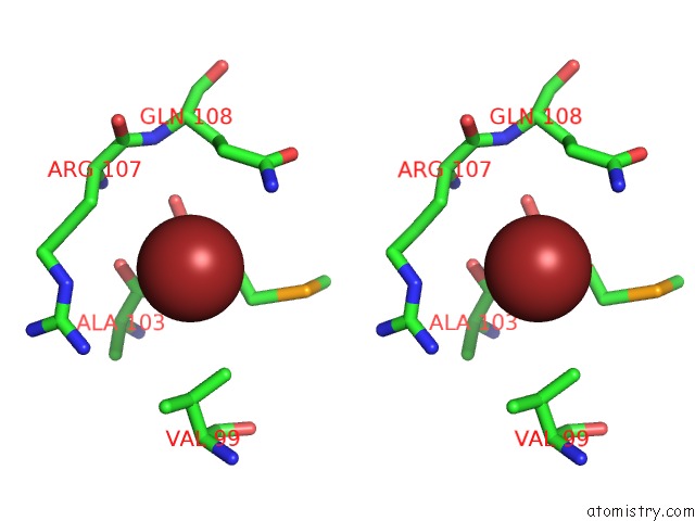
Stereo pair view
Mono view
Stereo pair view
A full contact list of Bromine with other atoms in the Br binding
site number 1 of Apoenzyme Crystal Structure of the Tuberculosis Serine/Threonine Kinase, Pkne within 5.0Å range:
|
Reference:
L.M.Gay,
H.L.Ng,
T.Alber.
A Conserved Dimer and Global Conformational Changes in the Structure of Apo-Pkne Ser/Thr Protein Kinase From Mycobacterium Tuberculosis. J.Mol.Biol. V. 360 409 2006.
ISSN: ISSN 0022-2836
PubMed: 16762364
DOI: 10.1016/J.JMB.2006.05.015
Page generated: Wed Jul 10 18:04:38 2024
ISSN: ISSN 0022-2836
PubMed: 16762364
DOI: 10.1016/J.JMB.2006.05.015
Last articles
Zn in 9J0NZn in 9J0O
Zn in 9J0P
Zn in 9FJX
Zn in 9EKB
Zn in 9C0F
Zn in 9CAH
Zn in 9CH0
Zn in 9CH3
Zn in 9CH1