Bromine »
PDB 2hc0-2jkl »
2jaw »
Bromine in PDB 2jaw: Crystal Structure of D41N Variant of Human Mitochondrial 5'(3')- Deoxyribonucleotidase (Mdn) in Complex with 5-Bromovinyldeoxyuridine 5'-Monophosphate
Protein crystallography data
The structure of Crystal Structure of D41N Variant of Human Mitochondrial 5'(3')- Deoxyribonucleotidase (Mdn) in Complex with 5-Bromovinyldeoxyuridine 5'-Monophosphate, PDB code: 2jaw
was solved by
K.Wallden,
B.Ruzzenente,
V.Bianchi,
P.Nordlund,
with X-Ray Crystallography technique. A brief refinement statistics is given in the table below:
| Resolution Low / High (Å) | 60.86 / 1.95 |
| Space group | P 43 21 2 |
| Cell size a, b, c (Å), α, β, γ (°) | 73.604, 73.604, 106.374, 90.00, 90.00, 90.00 |
| R / Rfree (%) | 18.5 / 21.6 |
Other elements in 2jaw:
The structure of Crystal Structure of D41N Variant of Human Mitochondrial 5'(3')- Deoxyribonucleotidase (Mdn) in Complex with 5-Bromovinyldeoxyuridine 5'-Monophosphate also contains other interesting chemical elements:
| Magnesium | (Mg) | 2 atoms |
Bromine Binding Sites:
The binding sites of Bromine atom in the Crystal Structure of D41N Variant of Human Mitochondrial 5'(3')- Deoxyribonucleotidase (Mdn) in Complex with 5-Bromovinyldeoxyuridine 5'-Monophosphate
(pdb code 2jaw). This binding sites where shown within
5.0 Angstroms radius around Bromine atom.
In total only one binding site of Bromine was determined in the Crystal Structure of D41N Variant of Human Mitochondrial 5'(3')- Deoxyribonucleotidase (Mdn) in Complex with 5-Bromovinyldeoxyuridine 5'-Monophosphate, PDB code: 2jaw:
In total only one binding site of Bromine was determined in the Crystal Structure of D41N Variant of Human Mitochondrial 5'(3')- Deoxyribonucleotidase (Mdn) in Complex with 5-Bromovinyldeoxyuridine 5'-Monophosphate, PDB code: 2jaw:
Bromine binding site 1 out of 1 in 2jaw
Go back to
Bromine binding site 1 out
of 1 in the Crystal Structure of D41N Variant of Human Mitochondrial 5'(3')- Deoxyribonucleotidase (Mdn) in Complex with 5-Bromovinyldeoxyuridine 5'-Monophosphate
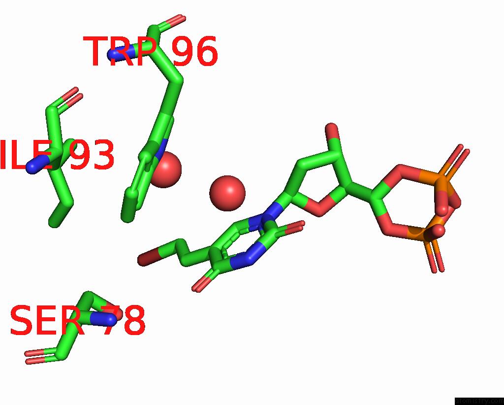
Mono view
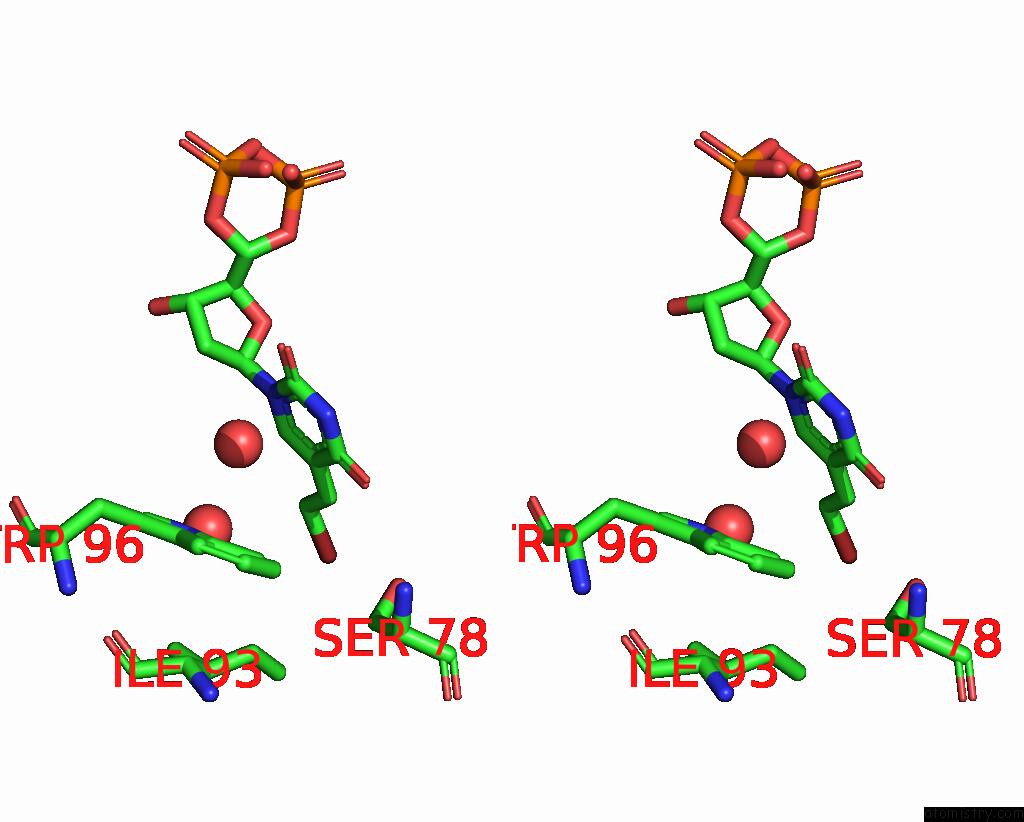
Stereo pair view
Mono view
Stereo pair view
A full contact list of Bromine with other atoms in the Br binding
site number 1 of Crystal Structure of D41N Variant of Human Mitochondrial 5'(3')- Deoxyribonucleotidase (Mdn) in Complex with 5-Bromovinyldeoxyuridine 5'-Monophosphate within 5.0Å range:
|
Reference:
K.Wallden,
A.Rinaldo-Matthis,
B.Ruzzenente,
C.Rampazzo,
V.Bianchi,
P.Nordlund.
Crystal Structures of Human and Murine Deoxyribonucleotidases: Insights Into Recognition of Substrates and Nucleotide Analogues. Biochemistry V. 46 13809 2007.
ISSN: ISSN 0006-2960
PubMed: 17985935
DOI: 10.1021/BI7014794
Page generated: Wed Jul 10 18:13:15 2024
ISSN: ISSN 0006-2960
PubMed: 17985935
DOI: 10.1021/BI7014794
Last articles
Zn in 9J0NZn in 9J0O
Zn in 9J0P
Zn in 9FJX
Zn in 9EKB
Zn in 9C0F
Zn in 9CAH
Zn in 9CH0
Zn in 9CH3
Zn in 9CH1