Bromine »
PDB 2jlt-2qbs »
2o2i »
Bromine in PDB 2o2i: Crystal Structure of Haloalkane Dehalogenase RV2579 From Mycobacterium Tuberculosis Complexed with 1,3-Propandiol
Enzymatic activity of Crystal Structure of Haloalkane Dehalogenase RV2579 From Mycobacterium Tuberculosis Complexed with 1,3-Propandiol
All present enzymatic activity of Crystal Structure of Haloalkane Dehalogenase RV2579 From Mycobacterium Tuberculosis Complexed with 1,3-Propandiol:
3.8.1.5;
3.8.1.5;
Protein crystallography data
The structure of Crystal Structure of Haloalkane Dehalogenase RV2579 From Mycobacterium Tuberculosis Complexed with 1,3-Propandiol, PDB code: 2o2i
was solved by
P.A.Mazumdar,
J.Hulecki,
M.M.Cherney,
C.R.Garen,
M.N.G.James,
Tbstructural Genomics Consortium (Tbsgc),
with X-Ray Crystallography technique. A brief refinement statistics is given in the table below:
| Resolution Low / High (Å) | 36.35 / 1.50 |
| Space group | C 2 2 21 |
| Cell size a, b, c (Å), α, β, γ (°) | 60.820, 65.190, 126.090, 90.00, 90.00, 90.00 |
| R / Rfree (%) | 14.4 / 17.6 |
Bromine Binding Sites:
The binding sites of Bromine atom in the Crystal Structure of Haloalkane Dehalogenase RV2579 From Mycobacterium Tuberculosis Complexed with 1,3-Propandiol
(pdb code 2o2i). This binding sites where shown within
5.0 Angstroms radius around Bromine atom.
In total only one binding site of Bromine was determined in the Crystal Structure of Haloalkane Dehalogenase RV2579 From Mycobacterium Tuberculosis Complexed with 1,3-Propandiol, PDB code: 2o2i:
In total only one binding site of Bromine was determined in the Crystal Structure of Haloalkane Dehalogenase RV2579 From Mycobacterium Tuberculosis Complexed with 1,3-Propandiol, PDB code: 2o2i:
Bromine binding site 1 out of 1 in 2o2i
Go back to
Bromine binding site 1 out
of 1 in the Crystal Structure of Haloalkane Dehalogenase RV2579 From Mycobacterium Tuberculosis Complexed with 1,3-Propandiol
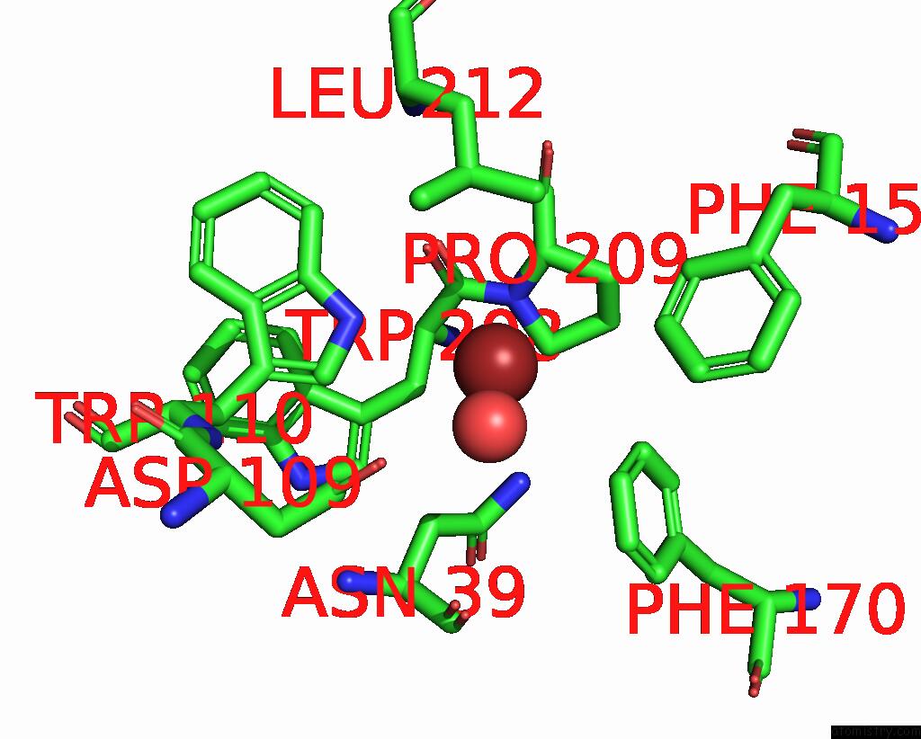
Mono view
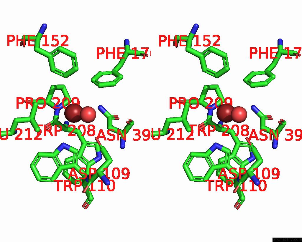
Stereo pair view
Mono view
Stereo pair view
A full contact list of Bromine with other atoms in the Br binding
site number 1 of Crystal Structure of Haloalkane Dehalogenase RV2579 From Mycobacterium Tuberculosis Complexed with 1,3-Propandiol within 5.0Å range:
|
Reference:
P.A.Mazumdar,
J.Hulecki,
M.M.Cherney,
C.R.Garen,
M.N.G.James.
Crystal Structure of Haloalkane Dehalogenase RV2579 From Mycobacterium Tuberculosis Complexed with 1,3-Propandiol To Be Published.
Page generated: Mon Jul 7 04:28:02 2025
Last articles
Br in 6C3UBr in 6CO5
Br in 6CNU
Br in 6CKE
Br in 6CCX
Br in 6CC9
Br in 6CJ9
Br in 6C6O
Br in 6CCM
Br in 6BXR