Bromine »
PDB 2qc6-2vo5 »
2r24 »
Bromine in PDB 2r24: Human Aldose Reductase Structure
Enzymatic activity of Human Aldose Reductase Structure
All present enzymatic activity of Human Aldose Reductase Structure:
1.1.1.21;
1.1.1.21;
Protein crystallography data
The structure of Human Aldose Reductase Structure, PDB code: 2r24
was solved by
M.P.Blakeley,
F.Ruiz,
R.Cachau,
I.Hazemann,
F.Meilleur,
A.Mitschler,
S.Ginell,
P.Afonine,
O.N.Ventura,
A.Cousido-Siah,
M.Haertlein,
A.Joachimiak,
D.Myles,
A.Podjarny,
with X-Ray Crystallography technique. A brief refinement statistics is given in the table below:
| Resolution Low / High (Å) | N/A / 1.75 |
| Space group | P 1 21 1 |
| Cell size a, b, c (Å), α, β, γ (°) | 50.066, 67.126, 47.862, 90.00, 92.41, 90.00 |
| R / Rfree (%) | 25.7 / 29.1 |
Other elements in 2r24:
The structure of Human Aldose Reductase Structure also contains other interesting chemical elements:
| Fluorine | (F) | 2 atoms |
Bromine Binding Sites:
The binding sites of Bromine atom in the Human Aldose Reductase Structure
(pdb code 2r24). This binding sites where shown within
5.0 Angstroms radius around Bromine atom.
In total only one binding site of Bromine was determined in the Human Aldose Reductase Structure, PDB code: 2r24:
In total only one binding site of Bromine was determined in the Human Aldose Reductase Structure, PDB code: 2r24:
Bromine binding site 1 out of 1 in 2r24
Go back to
Bromine binding site 1 out
of 1 in the Human Aldose Reductase Structure
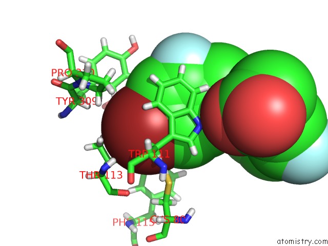
Mono view
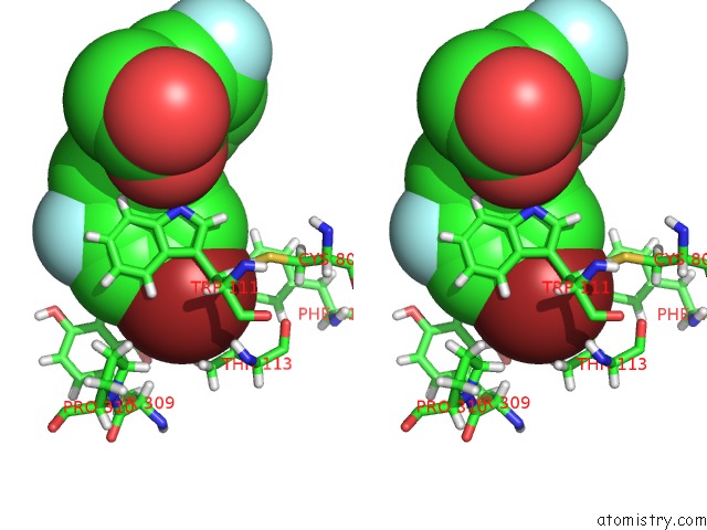
Stereo pair view
Mono view
Stereo pair view
A full contact list of Bromine with other atoms in the Br binding
site number 1 of Human Aldose Reductase Structure within 5.0Å range:
|
Reference:
M.P.Blakeley,
F.Ruiz,
R.Cachau,
I.Hazemann,
F.Meilleur,
A.Mitschler,
S.Ginell,
P.Afonine,
O.N.Ventura,
A.Cousido-Siah,
M.Haertlein,
A.Joachimiak,
D.Myles,
A.Podjarny.
Quantum Model of Catalysis Based on Mobile Proton Revealed By Subatomic X-Ray and Neutron Diffraction Studies of H-Aldose Reductase Proc.Natl.Acad.Sci.Usa V. 105 1844 2008.
ISSN: ISSN 0027-8424
PubMed: 18250329
DOI: 10.1073/PNAS.0711659105
Page generated: Mon Jul 7 04:38:24 2025
ISSN: ISSN 0027-8424
PubMed: 18250329
DOI: 10.1073/PNAS.0711659105
Last articles
F in 7M8OF in 7M8P
F in 7M7D
F in 7M63
F in 7M7N
F in 7M5Y
F in 7M5X
F in 7M5Z
F in 7M2N
F in 7M4V