Bromine »
PDB 2vot-2xo8 »
2wbk »
Bromine in PDB 2wbk: Structure of the Michaelis Complex of Beta-Mannosidase, MAN2A, Provides Insight Into the Conformational Itinerary of Mannoside Hydrolysis
Enzymatic activity of Structure of the Michaelis Complex of Beta-Mannosidase, MAN2A, Provides Insight Into the Conformational Itinerary of Mannoside Hydrolysis
All present enzymatic activity of Structure of the Michaelis Complex of Beta-Mannosidase, MAN2A, Provides Insight Into the Conformational Itinerary of Mannoside Hydrolysis:
3.2.1.25;
3.2.1.25;
Protein crystallography data
The structure of Structure of the Michaelis Complex of Beta-Mannosidase, MAN2A, Provides Insight Into the Conformational Itinerary of Mannoside Hydrolysis, PDB code: 2wbk
was solved by
W.A.Offen,
D.L.Zechel,
S.G.Withers,
H.J.Gilbert,
G.J.Davies,
with X-Ray Crystallography technique. A brief refinement statistics is given in the table below:
| Resolution Low / High (Å) | 47.84 / 2.10 |
| Space group | P 1 21 1 |
| Cell size a, b, c (Å), α, β, γ (°) | 91.447, 115.504, 97.705, 90.00, 115.99, 90.00 |
| R / Rfree (%) | 16.9 / 23.2 |
Other elements in 2wbk:
The structure of Structure of the Michaelis Complex of Beta-Mannosidase, MAN2A, Provides Insight Into the Conformational Itinerary of Mannoside Hydrolysis also contains other interesting chemical elements:
| Fluorine | (F) | 2 atoms |
| Chlorine | (Cl) | 7 atoms |
Bromine Binding Sites:
Pages:
>>> Page 1 <<< Page 2, Binding sites: 11 - 12;Binding sites:
The binding sites of Bromine atom in the Structure of the Michaelis Complex of Beta-Mannosidase, MAN2A, Provides Insight Into the Conformational Itinerary of Mannoside Hydrolysis (pdb code 2wbk). This binding sites where shown within 5.0 Angstroms radius around Bromine atom.In total 12 binding sites of Bromine where determined in the Structure of the Michaelis Complex of Beta-Mannosidase, MAN2A, Provides Insight Into the Conformational Itinerary of Mannoside Hydrolysis, PDB code: 2wbk:
Jump to Bromine binding site number: 1; 2; 3; 4; 5; 6; 7; 8; 9; 10;
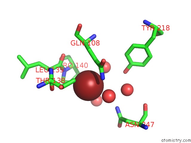
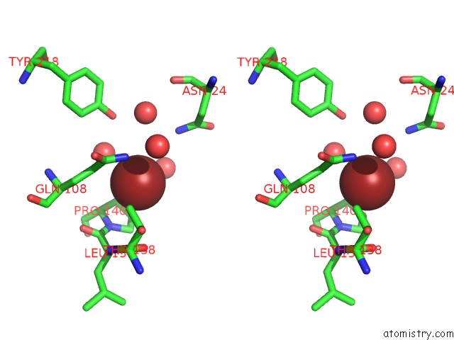
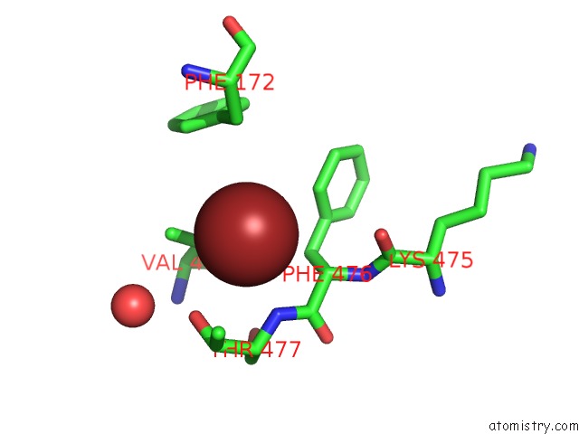
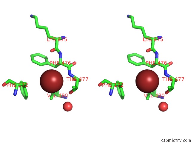
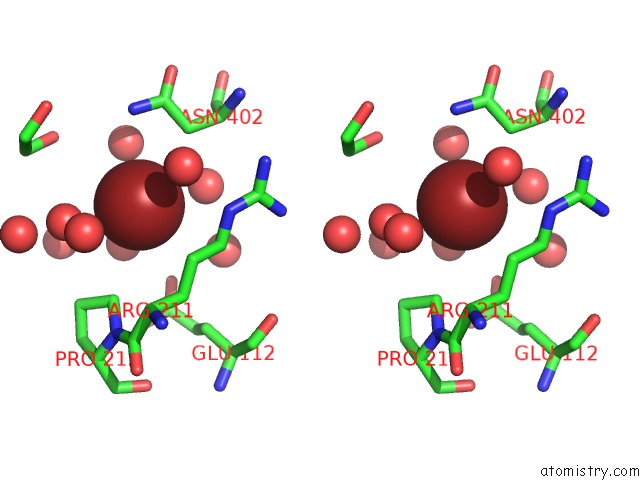
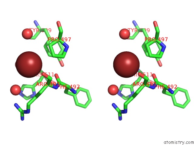
Bromine binding site 1 out of 12 in 2wbk
Go back to
Bromine binding site 1 out
of 12 in the Structure of the Michaelis Complex of Beta-Mannosidase, MAN2A, Provides Insight Into the Conformational Itinerary of Mannoside Hydrolysis

Mono view

Stereo pair view
Mono view
Stereo pair view
A full contact list of Bromine with other atoms in the Br binding
site number 1 of Structure of the Michaelis Complex of Beta-Mannosidase, MAN2A, Provides Insight Into the Conformational Itinerary of Mannoside Hydrolysis within 5.0Å range:
|
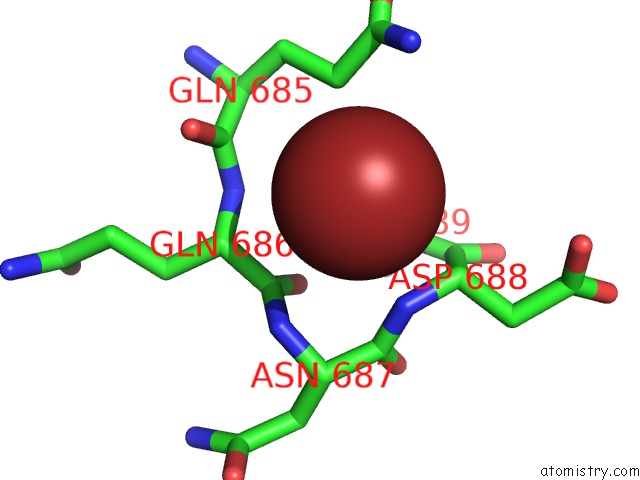
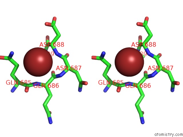
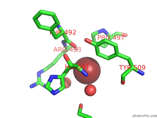
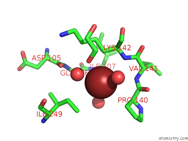
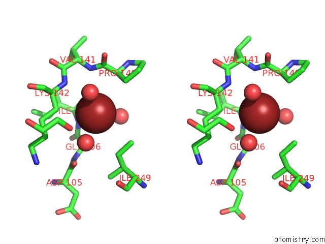
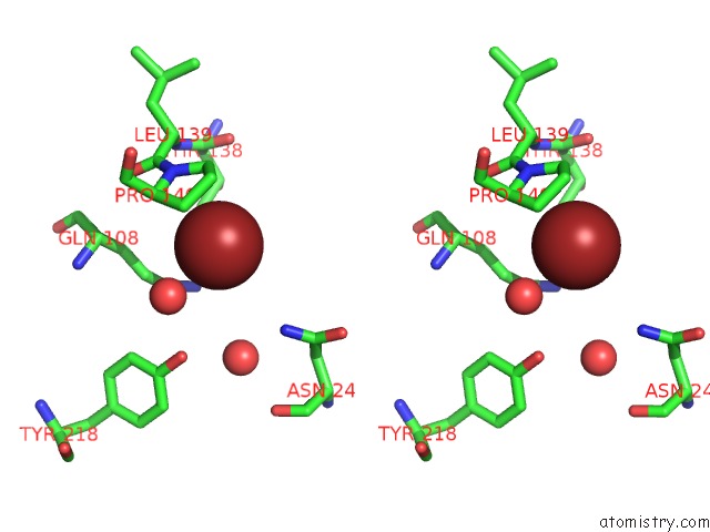
Bromine binding site 2 out of 12 in 2wbk
Go back to
Bromine binding site 2 out
of 12 in the Structure of the Michaelis Complex of Beta-Mannosidase, MAN2A, Provides Insight Into the Conformational Itinerary of Mannoside Hydrolysis
Mono view
Stereo pair view
Mono view
Stereo pair view
A full contact list of Bromine with other atoms in the Br binding
site number 2 of Structure of the Michaelis Complex of Beta-Mannosidase, MAN2A, Provides Insight Into the Conformational Itinerary of Mannoside Hydrolysis within 5.0Å range:
|
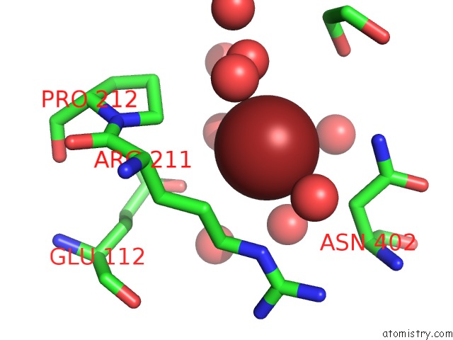
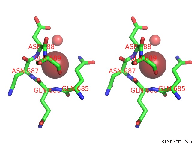
Bromine binding site 3 out of 12 in 2wbk
Go back to
Bromine binding site 3 out
of 12 in the Structure of the Michaelis Complex of Beta-Mannosidase, MAN2A, Provides Insight Into the Conformational Itinerary of Mannoside Hydrolysis
Mono view
Stereo pair view
Mono view
Stereo pair view
A full contact list of Bromine with other atoms in the Br binding
site number 3 of Structure of the Michaelis Complex of Beta-Mannosidase, MAN2A, Provides Insight Into the Conformational Itinerary of Mannoside Hydrolysis within 5.0Å range:
|
Bromine binding site 4 out of 12 in 2wbk
Go back to
Bromine binding site 4 out
of 12 in the Structure of the Michaelis Complex of Beta-Mannosidase, MAN2A, Provides Insight Into the Conformational Itinerary of Mannoside Hydrolysis
Mono view
Stereo pair view
Mono view
Stereo pair view
A full contact list of Bromine with other atoms in the Br binding
site number 4 of Structure of the Michaelis Complex of Beta-Mannosidase, MAN2A, Provides Insight Into the Conformational Itinerary of Mannoside Hydrolysis within 5.0Å range:
|
Bromine binding site 5 out of 12 in 2wbk
Go back to
Bromine binding site 5 out
of 12 in the Structure of the Michaelis Complex of Beta-Mannosidase, MAN2A, Provides Insight Into the Conformational Itinerary of Mannoside Hydrolysis
Mono view

Stereo pair view
Mono view
Stereo pair view
A full contact list of Bromine with other atoms in the Br binding
site number 5 of Structure of the Michaelis Complex of Beta-Mannosidase, MAN2A, Provides Insight Into the Conformational Itinerary of Mannoside Hydrolysis within 5.0Å range:
|
Bromine binding site 6 out of 12 in 2wbk
Go back to
Bromine binding site 6 out
of 12 in the Structure of the Michaelis Complex of Beta-Mannosidase, MAN2A, Provides Insight Into the Conformational Itinerary of Mannoside Hydrolysis
Mono view
Stereo pair view
Mono view
Stereo pair view
A full contact list of Bromine with other atoms in the Br binding
site number 6 of Structure of the Michaelis Complex of Beta-Mannosidase, MAN2A, Provides Insight Into the Conformational Itinerary of Mannoside Hydrolysis within 5.0Å range:
|
Bromine binding site 7 out of 12 in 2wbk
Go back to
Bromine binding site 7 out
of 12 in the Structure of the Michaelis Complex of Beta-Mannosidase, MAN2A, Provides Insight Into the Conformational Itinerary of Mannoside Hydrolysis
Mono view
Stereo pair view
Mono view
Stereo pair view
A full contact list of Bromine with other atoms in the Br binding
site number 7 of Structure of the Michaelis Complex of Beta-Mannosidase, MAN2A, Provides Insight Into the Conformational Itinerary of Mannoside Hydrolysis within 5.0Å range:
|
Bromine binding site 8 out of 12 in 2wbk
Go back to
Bromine binding site 8 out
of 12 in the Structure of the Michaelis Complex of Beta-Mannosidase, MAN2A, Provides Insight Into the Conformational Itinerary of Mannoside Hydrolysis

Mono view
Stereo pair view
Mono view
Stereo pair view
A full contact list of Bromine with other atoms in the Br binding
site number 8 of Structure of the Michaelis Complex of Beta-Mannosidase, MAN2A, Provides Insight Into the Conformational Itinerary of Mannoside Hydrolysis within 5.0Å range:
|
Bromine binding site 9 out of 12 in 2wbk
Go back to
Bromine binding site 9 out
of 12 in the Structure of the Michaelis Complex of Beta-Mannosidase, MAN2A, Provides Insight Into the Conformational Itinerary of Mannoside Hydrolysis

Mono view
Stereo pair view
Mono view
Stereo pair view
A full contact list of Bromine with other atoms in the Br binding
site number 9 of Structure of the Michaelis Complex of Beta-Mannosidase, MAN2A, Provides Insight Into the Conformational Itinerary of Mannoside Hydrolysis within 5.0Å range:
|
Bromine binding site 10 out of 12 in 2wbk
Go back to
Bromine binding site 10 out
of 12 in the Structure of the Michaelis Complex of Beta-Mannosidase, MAN2A, Provides Insight Into the Conformational Itinerary of Mannoside Hydrolysis

Mono view
Stereo pair view
Mono view
Stereo pair view
A full contact list of Bromine with other atoms in the Br binding
site number 10 of Structure of the Michaelis Complex of Beta-Mannosidase, MAN2A, Provides Insight Into the Conformational Itinerary of Mannoside Hydrolysis within 5.0Å range:
|
Reference:
W.A.Offen,
D.L.Zechel,
S.G.Withers,
H.J.Gilbert,
G.J.Davies.
Structure of the Michaelis Complex of Beta- Mannosidase, MAN2A, Provides Insight Into the Conformational Itinerary of Mannoside Hydrolysis. Cell(Cambridge,Mass.) V. 18 2484 2009.
ISSN: ISSN 0092-8674
PubMed: 19532864
DOI: 10.1039/B902240F
Page generated: Mon Jul 7 04:51:22 2025
ISSN: ISSN 0092-8674
PubMed: 19532864
DOI: 10.1039/B902240F
Last articles
Fe in 2YXOFe in 2YRS
Fe in 2YXC
Fe in 2YNM
Fe in 2YVJ
Fe in 2YP1
Fe in 2YU2
Fe in 2YU1
Fe in 2YQB
Fe in 2YOO