Bromine »
PDB 3bnr-3e01 »
3c51 »
Bromine in PDB 3c51: Crystal Structure of G Protein Coupled Receptor Kinase 1 Bound to Adp and Magnesium Chloride at 3.55A
Enzymatic activity of Crystal Structure of G Protein Coupled Receptor Kinase 1 Bound to Adp and Magnesium Chloride at 3.55A
All present enzymatic activity of Crystal Structure of G Protein Coupled Receptor Kinase 1 Bound to Adp and Magnesium Chloride at 3.55A:
2.7.11.14;
2.7.11.14;
Protein crystallography data
The structure of Crystal Structure of G Protein Coupled Receptor Kinase 1 Bound to Adp and Magnesium Chloride at 3.55A, PDB code: 3c51
was solved by
P.Singh,
J.J.G.Tesmer,
with X-Ray Crystallography technique. A brief refinement statistics is given in the table below:
| Resolution Low / High (Å) | 19.88 / 3.55 |
| Space group | P 21 21 21 |
| Cell size a, b, c (Å), α, β, γ (°) | 58.657, 92.527, 259.383, 90.00, 90.00, 90.00 |
| R / Rfree (%) | 28.1 / n/a |
Other elements in 3c51:
The structure of Crystal Structure of G Protein Coupled Receptor Kinase 1 Bound to Adp and Magnesium Chloride at 3.55A also contains other interesting chemical elements:
| Magnesium | (Mg) | 4 atoms |
Bromine Binding Sites:
The binding sites of Bromine atom in the Crystal Structure of G Protein Coupled Receptor Kinase 1 Bound to Adp and Magnesium Chloride at 3.55A
(pdb code 3c51). This binding sites where shown within
5.0 Angstroms radius around Bromine atom.
In total 5 binding sites of Bromine where determined in the Crystal Structure of G Protein Coupled Receptor Kinase 1 Bound to Adp and Magnesium Chloride at 3.55A, PDB code: 3c51:
Jump to Bromine binding site number: 1; 2; 3; 4; 5;
In total 5 binding sites of Bromine where determined in the Crystal Structure of G Protein Coupled Receptor Kinase 1 Bound to Adp and Magnesium Chloride at 3.55A, PDB code: 3c51:
Jump to Bromine binding site number: 1; 2; 3; 4; 5;
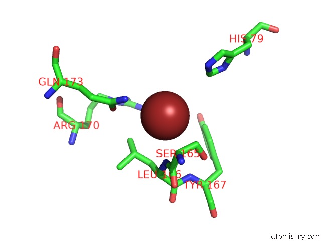
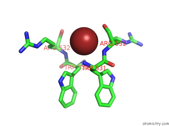
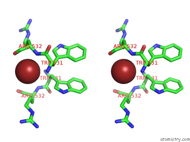
Bromine binding site 1 out of 5 in 3c51
Go back to
Bromine binding site 1 out
of 5 in the Crystal Structure of G Protein Coupled Receptor Kinase 1 Bound to Adp and Magnesium Chloride at 3.55A
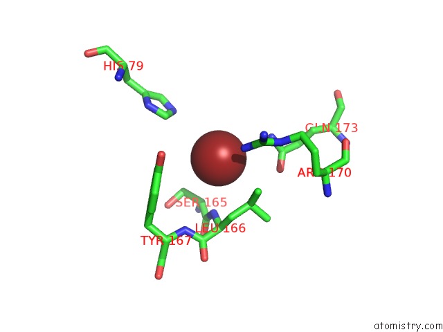
Mono view
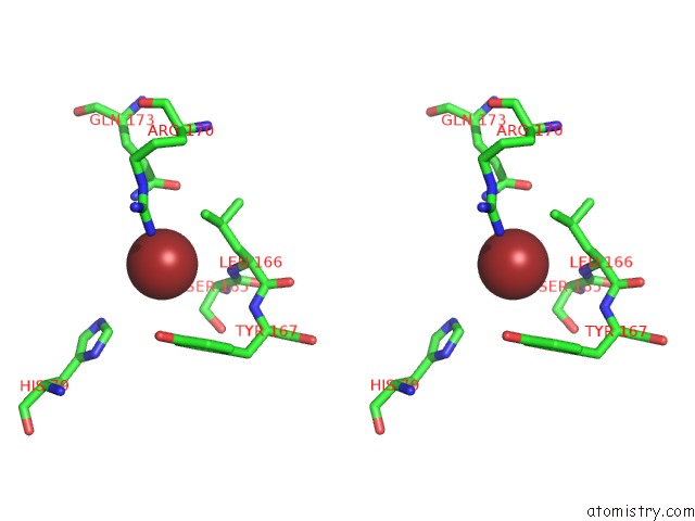
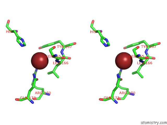
Stereo pair view
Mono view
Stereo pair view
A full contact list of Bromine with other atoms in the Br binding
site number 1 of Crystal Structure of G Protein Coupled Receptor Kinase 1 Bound to Adp and Magnesium Chloride at 3.55A within 5.0Å range:
|
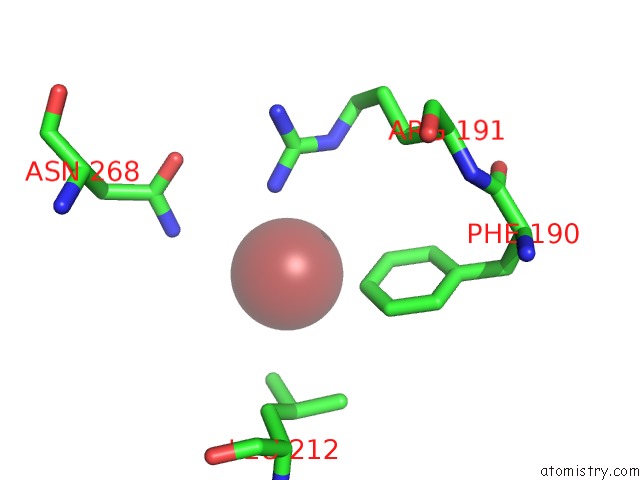
Bromine binding site 2 out of 5 in 3c51
Go back to
Bromine binding site 2 out
of 5 in the Crystal Structure of G Protein Coupled Receptor Kinase 1 Bound to Adp and Magnesium Chloride at 3.55A
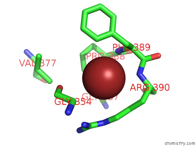
Mono view
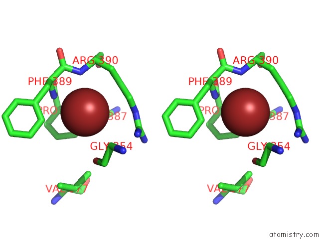
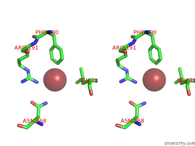
Stereo pair view
Mono view
Stereo pair view
A full contact list of Bromine with other atoms in the Br binding
site number 2 of Crystal Structure of G Protein Coupled Receptor Kinase 1 Bound to Adp and Magnesium Chloride at 3.55A within 5.0Å range:
|
Bromine binding site 3 out of 5 in 3c51
Go back to
Bromine binding site 3 out
of 5 in the Crystal Structure of G Protein Coupled Receptor Kinase 1 Bound to Adp and Magnesium Chloride at 3.55A
Mono view
Stereo pair view
Mono view
Stereo pair view
A full contact list of Bromine with other atoms in the Br binding
site number 3 of Crystal Structure of G Protein Coupled Receptor Kinase 1 Bound to Adp and Magnesium Chloride at 3.55A within 5.0Å range:
|
Bromine binding site 4 out of 5 in 3c51
Go back to
Bromine binding site 4 out
of 5 in the Crystal Structure of G Protein Coupled Receptor Kinase 1 Bound to Adp and Magnesium Chloride at 3.55A
Mono view
Stereo pair view
Mono view
Stereo pair view
A full contact list of Bromine with other atoms in the Br binding
site number 4 of Crystal Structure of G Protein Coupled Receptor Kinase 1 Bound to Adp and Magnesium Chloride at 3.55A within 5.0Å range:
|
Bromine binding site 5 out of 5 in 3c51
Go back to
Bromine binding site 5 out
of 5 in the Crystal Structure of G Protein Coupled Receptor Kinase 1 Bound to Adp and Magnesium Chloride at 3.55A
Mono view
Stereo pair view
Mono view
Stereo pair view
A full contact list of Bromine with other atoms in the Br binding
site number 5 of Crystal Structure of G Protein Coupled Receptor Kinase 1 Bound to Adp and Magnesium Chloride at 3.55A within 5.0Å range:
|
Reference:
P.Singh,
B.Wang,
T.Maeda,
K.Palczewski,
J.J.Tesmer.
Structures of Rhodopsin Kinase in Different Ligand States Reveal Key Elements Involved in G Protein-Coupled Receptor Kinase Activation. J.Biol.Chem. V. 283 14053 2008.
ISSN: ISSN 0021-9258
PubMed: 18339619
DOI: 10.1074/JBC.M708974200
Page generated: Wed Jul 10 19:11:50 2024
ISSN: ISSN 0021-9258
PubMed: 18339619
DOI: 10.1074/JBC.M708974200
Last articles
Zn in 9MJ5Zn in 9HNW
Zn in 9G0L
Zn in 9FNE
Zn in 9DZN
Zn in 9E0I
Zn in 9D32
Zn in 9DAK
Zn in 8ZXC
Zn in 8ZUF