Bromine »
PDB 3e0q-3gud »
3f59 »
Bromine in PDB 3f59: Crystal Structure of ZU5-Ank, the Spectrin Binding Region of Human Erythroid Ankyrin
Protein crystallography data
The structure of Crystal Structure of ZU5-Ank, the Spectrin Binding Region of Human Erythroid Ankyrin, PDB code: 3f59
was solved by
J.J.Ipsaro,
L.Huang,
A.Mondragon,
with X-Ray Crystallography technique. A brief refinement statistics is given in the table below:
| Resolution Low / High (Å) | 36.91 / 2.00 |
| Space group | P 1 21 1 |
| Cell size a, b, c (Å), α, β, γ (°) | 38.040, 204.110, 43.070, 90.00, 113.14, 90.00 |
| R / Rfree (%) | 21.6 / 26.4 |
Bromine Binding Sites:
Pages:
>>> Page 1 <<< Page 2, Binding sites: 11 - 18;Binding sites:
The binding sites of Bromine atom in the Crystal Structure of ZU5-Ank, the Spectrin Binding Region of Human Erythroid Ankyrin (pdb code 3f59). This binding sites where shown within 5.0 Angstroms radius around Bromine atom.In total 18 binding sites of Bromine where determined in the Crystal Structure of ZU5-Ank, the Spectrin Binding Region of Human Erythroid Ankyrin, PDB code: 3f59:
Jump to Bromine binding site number: 1; 2; 3; 4; 5; 6; 7; 8; 9; 10;
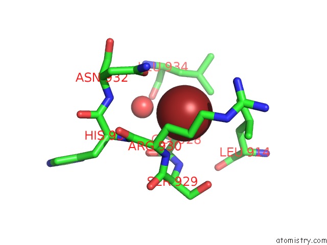
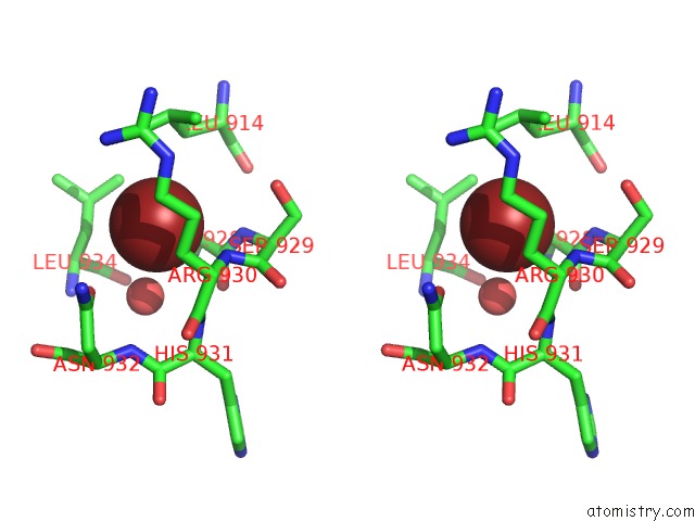
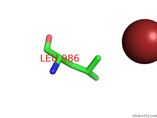
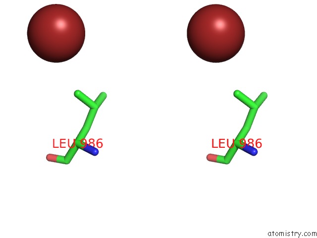
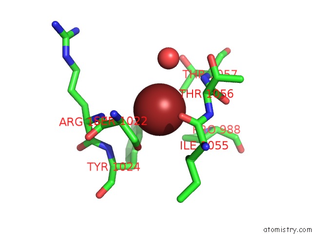
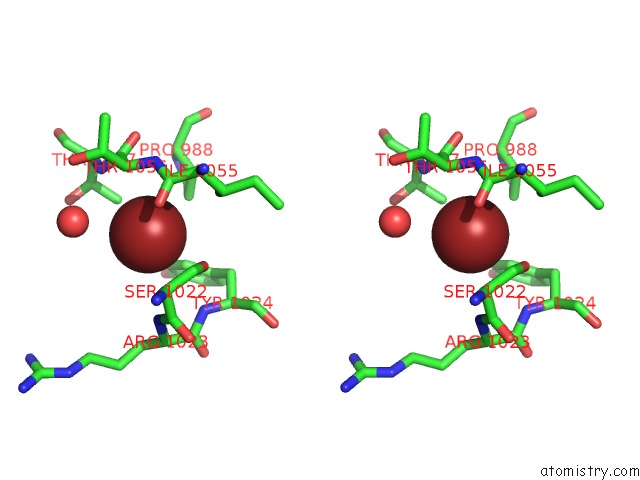
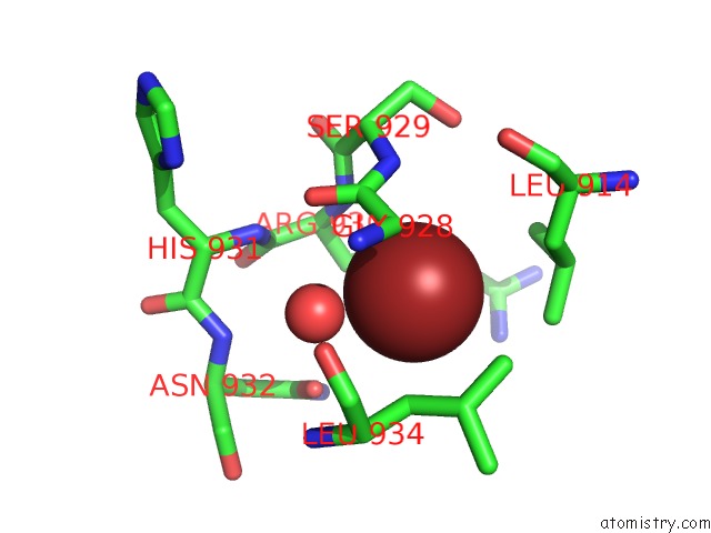
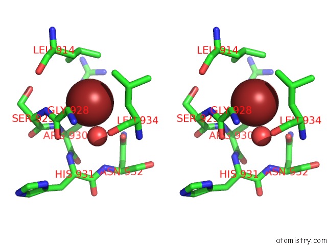
Bromine binding site 1 out of 18 in 3f59
Go back to
Bromine binding site 1 out
of 18 in the Crystal Structure of ZU5-Ank, the Spectrin Binding Region of Human Erythroid Ankyrin
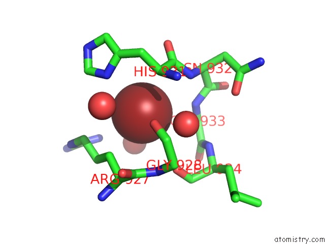
Mono view
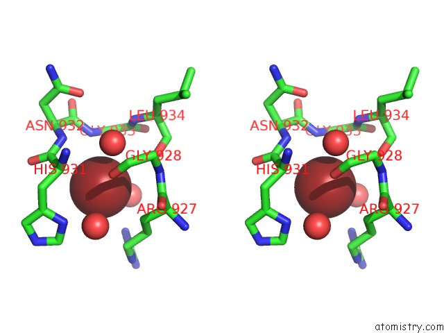
Stereo pair view
Mono view
Stereo pair view
A full contact list of Bromine with other atoms in the Br binding
site number 1 of Crystal Structure of ZU5-Ank, the Spectrin Binding Region of Human Erythroid Ankyrin within 5.0Å range:
|
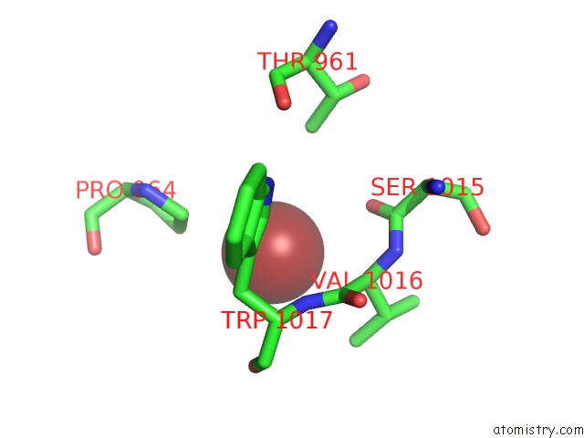
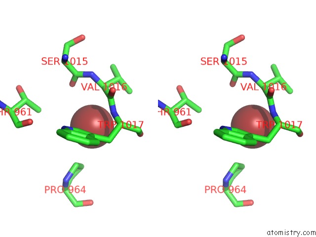
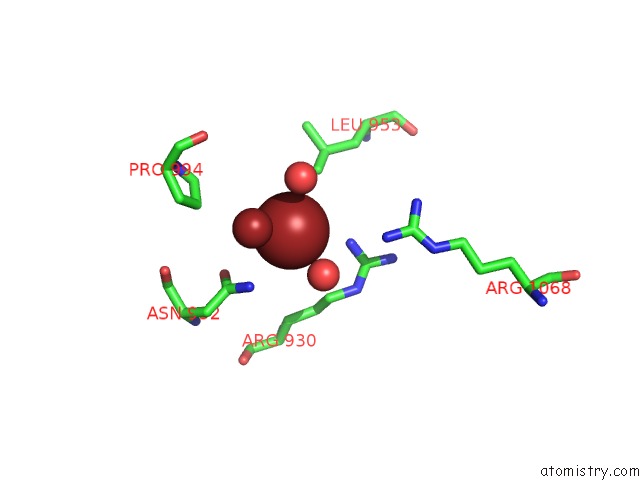
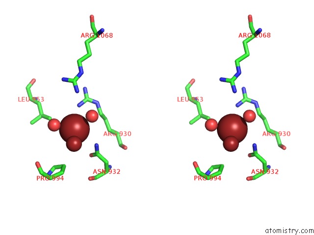
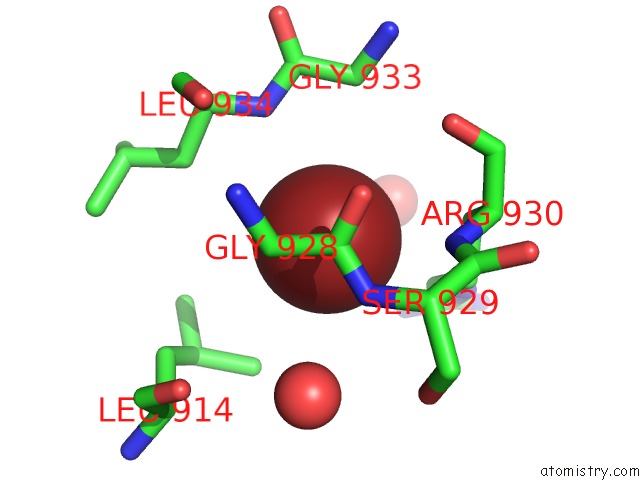
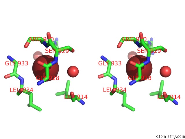
Bromine binding site 2 out of 18 in 3f59
Go back to
Bromine binding site 2 out
of 18 in the Crystal Structure of ZU5-Ank, the Spectrin Binding Region of Human Erythroid Ankyrin
Mono view
Stereo pair view
Mono view
Stereo pair view
A full contact list of Bromine with other atoms in the Br binding
site number 2 of Crystal Structure of ZU5-Ank, the Spectrin Binding Region of Human Erythroid Ankyrin within 5.0Å range:
|
Bromine binding site 3 out of 18 in 3f59
Go back to
Bromine binding site 3 out
of 18 in the Crystal Structure of ZU5-Ank, the Spectrin Binding Region of Human Erythroid Ankyrin
Mono view
Stereo pair view
Mono view
Stereo pair view
A full contact list of Bromine with other atoms in the Br binding
site number 3 of Crystal Structure of ZU5-Ank, the Spectrin Binding Region of Human Erythroid Ankyrin within 5.0Å range:
|
Bromine binding site 4 out of 18 in 3f59
Go back to
Bromine binding site 4 out
of 18 in the Crystal Structure of ZU5-Ank, the Spectrin Binding Region of Human Erythroid Ankyrin
Mono view
Stereo pair view
Mono view
Stereo pair view
| A full contact list of Bromine with other atoms in the Br binding site number 4 of Crystal Structure of ZU5-Ank, the Spectrin Binding Region of Human Erythroid Ankyrin within 5.0Å range: |
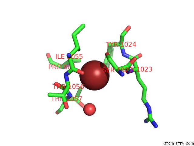
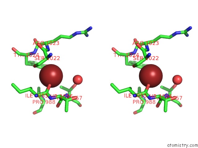
Bromine binding site 5 out of 18 in 3f59
Go back to
Bromine binding site 5 out
of 18 in the Crystal Structure of ZU5-Ank, the Spectrin Binding Region of Human Erythroid Ankyrin
Mono view
Stereo pair view
Mono view
Stereo pair view
A full contact list of Bromine with other atoms in the Br binding
site number 5 of Crystal Structure of ZU5-Ank, the Spectrin Binding Region of Human Erythroid Ankyrin within 5.0Å range:
|
Bromine binding site 6 out of 18 in 3f59
Go back to
Bromine binding site 6 out
of 18 in the Crystal Structure of ZU5-Ank, the Spectrin Binding Region of Human Erythroid Ankyrin
Mono view
Stereo pair view
Mono view
Stereo pair view
A full contact list of Bromine with other atoms in the Br binding
site number 6 of Crystal Structure of ZU5-Ank, the Spectrin Binding Region of Human Erythroid Ankyrin within 5.0Å range:
|
Bromine binding site 7 out of 18 in 3f59
Go back to
Bromine binding site 7 out
of 18 in the Crystal Structure of ZU5-Ank, the Spectrin Binding Region of Human Erythroid Ankyrin
Mono view
Stereo pair view
Mono view
Stereo pair view
A full contact list of Bromine with other atoms in the Br binding
site number 7 of Crystal Structure of ZU5-Ank, the Spectrin Binding Region of Human Erythroid Ankyrin within 5.0Å range:
|
Bromine binding site 8 out of 18 in 3f59
Go back to
Bromine binding site 8 out
of 18 in the Crystal Structure of ZU5-Ank, the Spectrin Binding Region of Human Erythroid Ankyrin
Mono view
Stereo pair view
Mono view
Stereo pair view
A full contact list of Bromine with other atoms in the Br binding
site number 8 of Crystal Structure of ZU5-Ank, the Spectrin Binding Region of Human Erythroid Ankyrin within 5.0Å range:
|
Bromine binding site 9 out of 18 in 3f59
Go back to
Bromine binding site 9 out
of 18 in the Crystal Structure of ZU5-Ank, the Spectrin Binding Region of Human Erythroid Ankyrin
Mono view
Stereo pair view
Mono view
Stereo pair view
A full contact list of Bromine with other atoms in the Br binding
site number 9 of Crystal Structure of ZU5-Ank, the Spectrin Binding Region of Human Erythroid Ankyrin within 5.0Å range:
|
Bromine binding site 10 out of 18 in 3f59
Go back to
Bromine binding site 10 out
of 18 in the Crystal Structure of ZU5-Ank, the Spectrin Binding Region of Human Erythroid Ankyrin
Mono view
Stereo pair view
Mono view
Stereo pair view
A full contact list of Bromine with other atoms in the Br binding
site number 10 of Crystal Structure of ZU5-Ank, the Spectrin Binding Region of Human Erythroid Ankyrin within 5.0Å range:
|
Reference:
J.J.Ipsaro,
L.Huang,
A.Mondragon.
Structures of the Spectrin-Ankyrin Interaction Binding Domains. Blood V. 113 5385 2009.
ISSN: ISSN 0006-4971
PubMed: 19141864
DOI: 10.1182/BLOOD-2008-10-184358
Page generated: Mon Jul 7 05:17:58 2025
ISSN: ISSN 0006-4971
PubMed: 19141864
DOI: 10.1182/BLOOD-2008-10-184358
Last articles
Cl in 3MBHCl in 3MBY
Cl in 3MB4
Cl in 3MBJ
Cl in 3M9F
Cl in 3MBD
Cl in 3MAA
Cl in 3MAF
Cl in 3M8O
Cl in 3M6Y