Bromine »
PDB 3gw6-3jtk »
3ivn »
Bromine in PDB 3ivn: Structure of the U65C Mutant A-Riboswitch Aptamer From the Bacillus Subtilis Pbue Operon
Protein crystallography data
The structure of Structure of the U65C Mutant A-Riboswitch Aptamer From the Bacillus Subtilis Pbue Operon, PDB code: 3ivn
was solved by
V.Delfosse,
P.Dagenais,
D.Chausse,
G.Di Tomasso,
P.Legault,
with X-Ray Crystallography technique. A brief refinement statistics is given in the table below:
| Resolution Low / High (Å) | 26.04 / 2.80 |
| Space group | C 1 2 1 |
| Cell size a, b, c (Å), α, β, γ (°) | 124.930, 46.330, 87.240, 90.00, 120.04, 90.00 |
| R / Rfree (%) | 22.7 / 27.7 |
Other elements in 3ivn:
The structure of Structure of the U65C Mutant A-Riboswitch Aptamer From the Bacillus Subtilis Pbue Operon also contains other interesting chemical elements:
| Magnesium | (Mg) | 9 atoms |
Bromine Binding Sites:
The binding sites of Bromine atom in the Structure of the U65C Mutant A-Riboswitch Aptamer From the Bacillus Subtilis Pbue Operon
(pdb code 3ivn). This binding sites where shown within
5.0 Angstroms radius around Bromine atom.
In total 2 binding sites of Bromine where determined in the Structure of the U65C Mutant A-Riboswitch Aptamer From the Bacillus Subtilis Pbue Operon, PDB code: 3ivn:
Jump to Bromine binding site number: 1; 2;
In total 2 binding sites of Bromine where determined in the Structure of the U65C Mutant A-Riboswitch Aptamer From the Bacillus Subtilis Pbue Operon, PDB code: 3ivn:
Jump to Bromine binding site number: 1; 2;
Bromine binding site 1 out of 2 in 3ivn
Go back to
Bromine binding site 1 out
of 2 in the Structure of the U65C Mutant A-Riboswitch Aptamer From the Bacillus Subtilis Pbue Operon
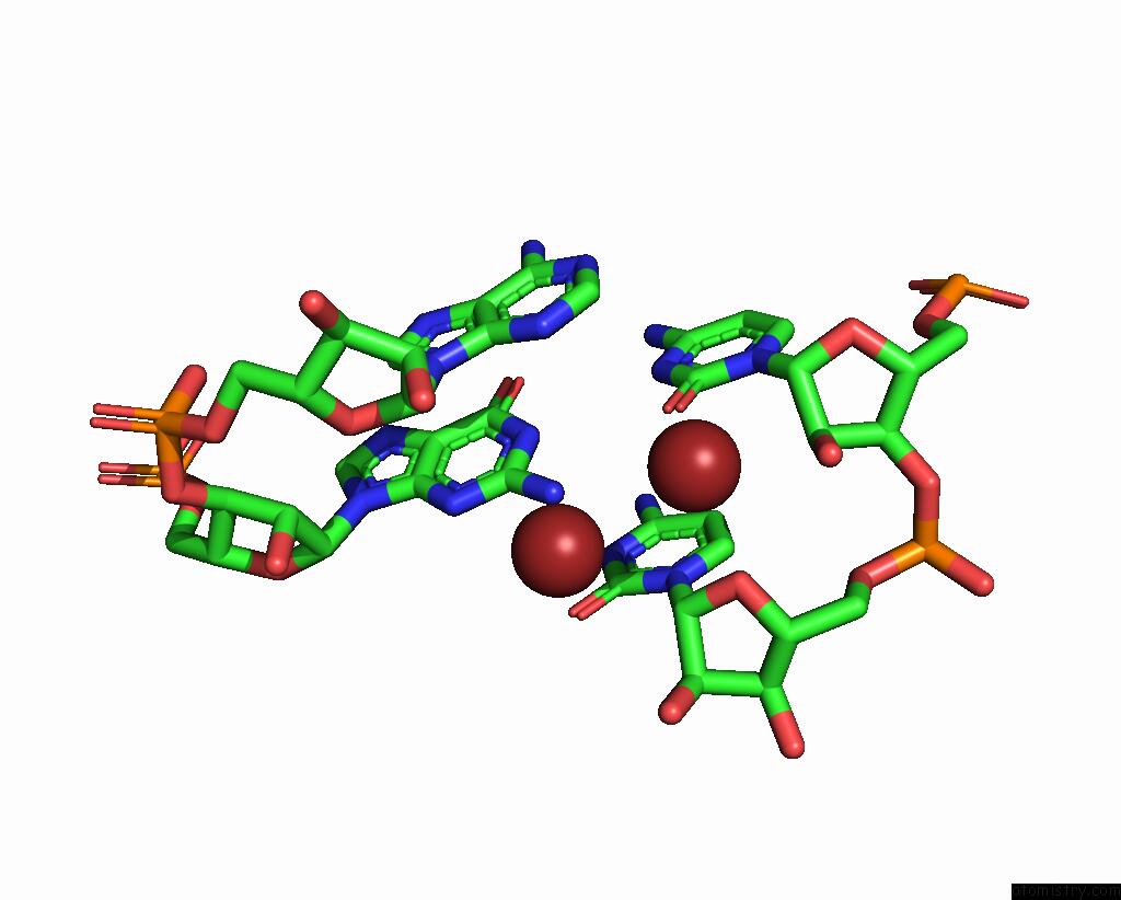
Mono view
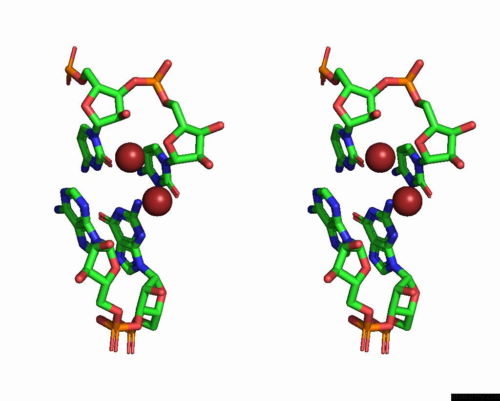
Stereo pair view
Mono view
Stereo pair view
A full contact list of Bromine with other atoms in the Br binding
site number 1 of Structure of the U65C Mutant A-Riboswitch Aptamer From the Bacillus Subtilis Pbue Operon within 5.0Å range:
|
Bromine binding site 2 out of 2 in 3ivn
Go back to
Bromine binding site 2 out
of 2 in the Structure of the U65C Mutant A-Riboswitch Aptamer From the Bacillus Subtilis Pbue Operon
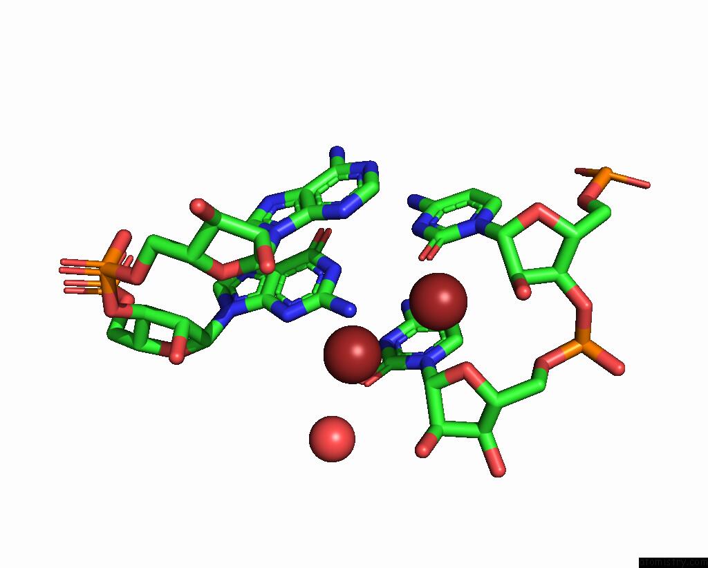
Mono view
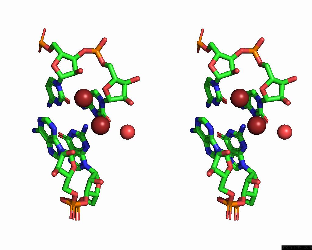
Stereo pair view
Mono view
Stereo pair view
A full contact list of Bromine with other atoms in the Br binding
site number 2 of Structure of the U65C Mutant A-Riboswitch Aptamer From the Bacillus Subtilis Pbue Operon within 5.0Å range:
|
Reference:
V.Delfosse,
P.Bouchard,
E.Bonneau,
P.Dagenais,
J.F.Lemay,
D.A.Lafontaine,
P.Legault.
Riboswitch Structure: An Internal Residue Mimicking the Purine Ligand. Nucleic Acids Res. V. 38 2057 2010.
ISSN: ISSN 0305-1048
PubMed: 20022916
DOI: 10.1093/NAR/GKP1080
Page generated: Mon Jul 7 05:28:01 2025
ISSN: ISSN 0305-1048
PubMed: 20022916
DOI: 10.1093/NAR/GKP1080
Last articles
F in 7G2GF in 7G1H
F in 7G2F
F in 7G1I
F in 7G2E
F in 7G1K
F in 7G1P
F in 7G0I
F in 7G05
F in 7G18