Bromine »
PDB 3ok5-3r1e »
3qyw »
Bromine in PDB 3qyw: Crystal Structure of ERK2 in Complex with An Inhibitor
Enzymatic activity of Crystal Structure of ERK2 in Complex with An Inhibitor
All present enzymatic activity of Crystal Structure of ERK2 in Complex with An Inhibitor:
2.7.11.24;
2.7.11.24;
Protein crystallography data
The structure of Crystal Structure of ERK2 in Complex with An Inhibitor, PDB code: 3qyw
was solved by
M.Gelin,
S.Pochet,
F.Hoh,
M.Pirochi,
J.-F.Guichou,
J.-L.Ferrer,
G.Labesse,
with X-Ray Crystallography technique. A brief refinement statistics is given in the table below:
| Resolution Low / High (Å) | 36.98 / 1.50 |
| Space group | P 1 21 1 |
| Cell size a, b, c (Å), α, β, γ (°) | 49.067, 70.254, 60.168, 90.00, 108.72, 90.00 |
| R / Rfree (%) | 14.8 / 18.8 |
Bromine Binding Sites:
The binding sites of Bromine atom in the Crystal Structure of ERK2 in Complex with An Inhibitor
(pdb code 3qyw). This binding sites where shown within
5.0 Angstroms radius around Bromine atom.
In total only one binding site of Bromine was determined in the Crystal Structure of ERK2 in Complex with An Inhibitor, PDB code: 3qyw:
In total only one binding site of Bromine was determined in the Crystal Structure of ERK2 in Complex with An Inhibitor, PDB code: 3qyw:
Bromine binding site 1 out of 1 in 3qyw
Go back to
Bromine binding site 1 out
of 1 in the Crystal Structure of ERK2 in Complex with An Inhibitor
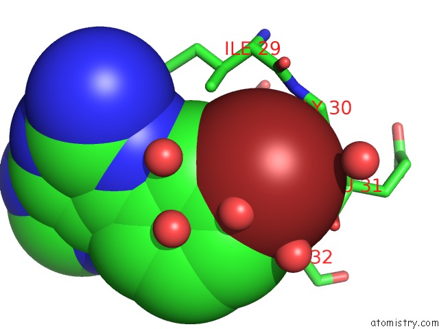
Mono view
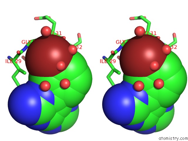
Stereo pair view
Mono view
Stereo pair view
A full contact list of Bromine with other atoms in the Br binding
site number 1 of Crystal Structure of ERK2 in Complex with An Inhibitor within 5.0Å range:
|
Reference:
A.Le Maire,
M.Gelin,
S.Pochet,
F.Hoh,
M.Pirocchi,
J.F.Guichou,
J.L.Ferrer,
G.Labesse.
In-Plate Protein Crystallization, in Situ Ligand Soaking and X-Ray Diffraction. Acta Crystallogr.,Sect.D V. 67 747 2011.
ISSN: ISSN 0907-4449
PubMed: 21904027
DOI: 10.1107/S0907444911023249
Page generated: Mon Jul 7 05:51:31 2025
ISSN: ISSN 0907-4449
PubMed: 21904027
DOI: 10.1107/S0907444911023249
Last articles
Br in 8KI3Br in 8K4H
Br in 8KHF
Br in 8KH6
Br in 8KE4
Br in 8KE1
Br in 8K2M
Br in 8K9L
Br in 8K6D
Br in 8JV5