Bromine »
PDB 3r3x-3tv4 »
3stv »
Bromine in PDB 3stv: Crystal Structure of Tomato Methylketone Synthase I Complexed with 3- Hydroxyoctanoate
Protein crystallography data
The structure of Crystal Structure of Tomato Methylketone Synthase I Complexed with 3- Hydroxyoctanoate, PDB code: 3stv
was solved by
M.E.Auldridge,
M.B.Austin,
J.P.Noel,
with X-Ray Crystallography technique. A brief refinement statistics is given in the table below:
| Resolution Low / High (Å) | 19.60 / 2.20 |
| Space group | P 1 21 1 |
| Cell size a, b, c (Å), α, β, γ (°) | 48.038, 92.380, 60.596, 90.00, 97.56, 90.00 |
| R / Rfree (%) | 20.2 / 23.6 |
Bromine Binding Sites:
The binding sites of Bromine atom in the Crystal Structure of Tomato Methylketone Synthase I Complexed with 3- Hydroxyoctanoate
(pdb code 3stv). This binding sites where shown within
5.0 Angstroms radius around Bromine atom.
In total only one binding site of Bromine was determined in the Crystal Structure of Tomato Methylketone Synthase I Complexed with 3- Hydroxyoctanoate, PDB code: 3stv:
In total only one binding site of Bromine was determined in the Crystal Structure of Tomato Methylketone Synthase I Complexed with 3- Hydroxyoctanoate, PDB code: 3stv:
Bromine binding site 1 out of 1 in 3stv
Go back to
Bromine binding site 1 out
of 1 in the Crystal Structure of Tomato Methylketone Synthase I Complexed with 3- Hydroxyoctanoate
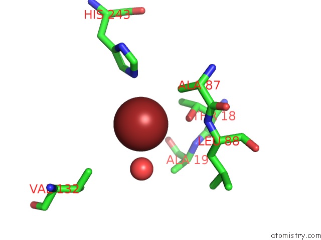
Mono view
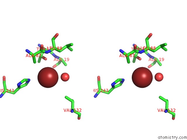
Stereo pair view
Mono view
Stereo pair view
A full contact list of Bromine with other atoms in the Br binding
site number 1 of Crystal Structure of Tomato Methylketone Synthase I Complexed with 3- Hydroxyoctanoate within 5.0Å range:
|
Reference:
M.E.Auldridge,
Y.Guo,
M.B.Austin,
J.Ramsey,
E.Fridman,
E.Pichersky,
J.P.Noel.
Emergent Decarboxylase Activity and Attenuation of Alpha/Beta-Hydrolase Activity During the Evolution of Methylketone Biosynthesis in Tomato. Plant Cell V. 24 1596 2012.
ISSN: ISSN 1040-4651
PubMed: 22523203
DOI: 10.1105/TPC.111.093997
Page generated: Wed Jul 10 20:21:51 2024
ISSN: ISSN 1040-4651
PubMed: 22523203
DOI: 10.1105/TPC.111.093997
Last articles
Zn in 9MJ5Zn in 9HNW
Zn in 9G0L
Zn in 9FNE
Zn in 9DZN
Zn in 9E0I
Zn in 9D32
Zn in 9DAK
Zn in 8ZXC
Zn in 8ZUF