Bromine »
PDB 4mye-4p5b »
4ohu »
Bromine in PDB 4ohu: Crystal Structure of Mycobacterium Tuberculosis Inha in Complex with Inhibitor PT92
Enzymatic activity of Crystal Structure of Mycobacterium Tuberculosis Inha in Complex with Inhibitor PT92
All present enzymatic activity of Crystal Structure of Mycobacterium Tuberculosis Inha in Complex with Inhibitor PT92:
1.3.1.9;
1.3.1.9;
Protein crystallography data
The structure of Crystal Structure of Mycobacterium Tuberculosis Inha in Complex with Inhibitor PT92, PDB code: 4ohu
was solved by
H.J.Li,
P.Pan,
C.T.Lai,
N.Liu,
W.Yu,
M.Garcia-Diaz,
C.Simmerling,
P.J.Tonge,
with X-Ray Crystallography technique. A brief refinement statistics is given in the table below:
| Resolution Low / High (Å) | 28.63 / 1.60 |
| Space group | P 21 21 21 |
| Cell size a, b, c (Å), α, β, γ (°) | 72.845, 90.486, 161.825, 90.00, 90.00, 90.00 |
| R / Rfree (%) | 17.1 / 18.8 |
Bromine Binding Sites:
The binding sites of Bromine atom in the Crystal Structure of Mycobacterium Tuberculosis Inha in Complex with Inhibitor PT92
(pdb code 4ohu). This binding sites where shown within
5.0 Angstroms radius around Bromine atom.
In total 4 binding sites of Bromine where determined in the Crystal Structure of Mycobacterium Tuberculosis Inha in Complex with Inhibitor PT92, PDB code: 4ohu:
Jump to Bromine binding site number: 1; 2; 3; 4;
In total 4 binding sites of Bromine where determined in the Crystal Structure of Mycobacterium Tuberculosis Inha in Complex with Inhibitor PT92, PDB code: 4ohu:
Jump to Bromine binding site number: 1; 2; 3; 4;
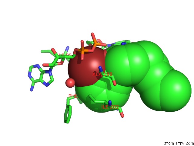
Bromine binding site 1 out of 4 in 4ohu
Go back to
Bromine binding site 1 out
of 4 in the Crystal Structure of Mycobacterium Tuberculosis Inha in Complex with Inhibitor PT92
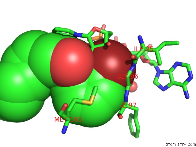
Mono view
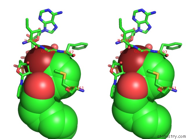
Stereo pair view
Mono view
Stereo pair view
A full contact list of Bromine with other atoms in the Br binding
site number 1 of Crystal Structure of Mycobacterium Tuberculosis Inha in Complex with Inhibitor PT92 within 5.0Å range:
|
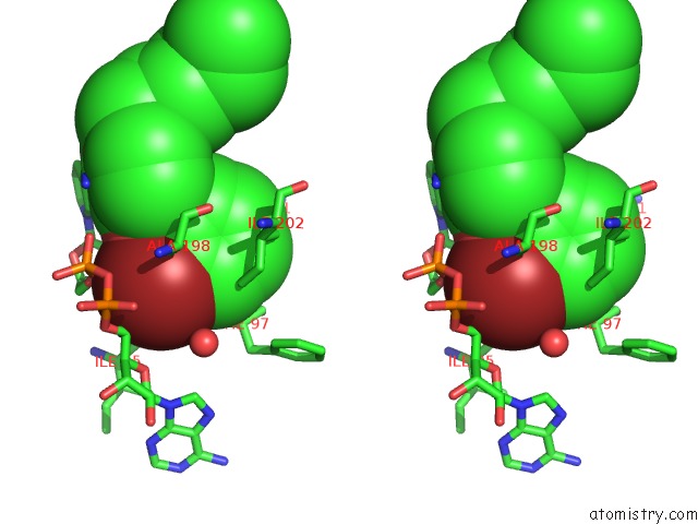
Bromine binding site 2 out of 4 in 4ohu
Go back to
Bromine binding site 2 out
of 4 in the Crystal Structure of Mycobacterium Tuberculosis Inha in Complex with Inhibitor PT92
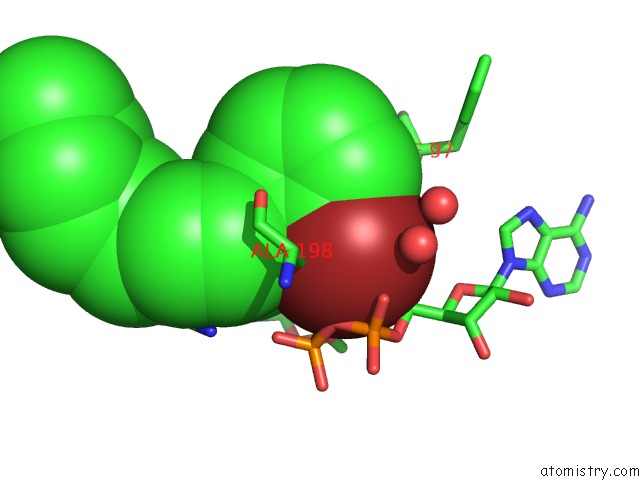
Mono view
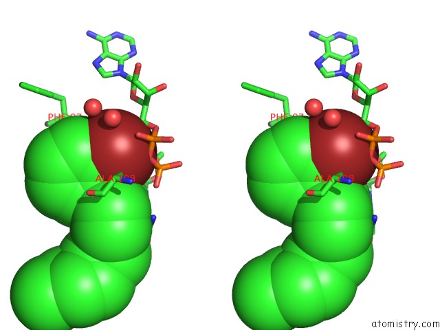
Stereo pair view
Mono view
Stereo pair view
A full contact list of Bromine with other atoms in the Br binding
site number 2 of Crystal Structure of Mycobacterium Tuberculosis Inha in Complex with Inhibitor PT92 within 5.0Å range:
|
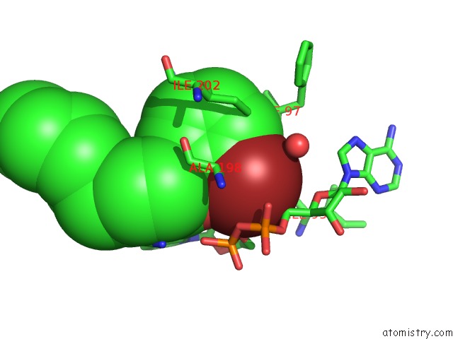
Bromine binding site 3 out of 4 in 4ohu
Go back to
Bromine binding site 3 out
of 4 in the Crystal Structure of Mycobacterium Tuberculosis Inha in Complex with Inhibitor PT92
Mono view
Stereo pair view
Mono view
Stereo pair view
A full contact list of Bromine with other atoms in the Br binding
site number 3 of Crystal Structure of Mycobacterium Tuberculosis Inha in Complex with Inhibitor PT92 within 5.0Å range:
|
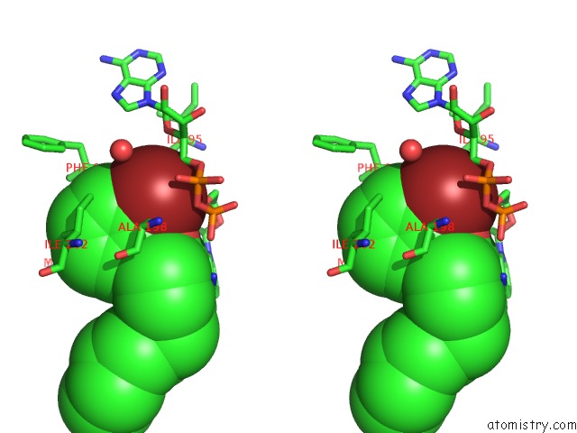
Bromine binding site 4 out of 4 in 4ohu
Go back to
Bromine binding site 4 out
of 4 in the Crystal Structure of Mycobacterium Tuberculosis Inha in Complex with Inhibitor PT92
Mono view
Stereo pair view
Mono view
Stereo pair view
A full contact list of Bromine with other atoms in the Br binding
site number 4 of Crystal Structure of Mycobacterium Tuberculosis Inha in Complex with Inhibitor PT92 within 5.0Å range:
|
Reference:
H.J.Li,
C.T.Lai,
P.Pan,
W.Yu,
N.Liu,
G.R.Bommineni,
M.Garcia-Diaz,
C.Simmerling,
P.J.Tonge.
A Structural and Energetic Model For the Slow-Onset Inhibition of the Mycobacterium Tuberculosis Enoyl-Acp Reductase Inha. Acs Chem.Biol. V. 9 986 2014.
ISSN: ISSN 1554-8929
PubMed: 24527857
DOI: 10.1021/CB400896G
Page generated: Wed Jul 10 22:11:19 2024
ISSN: ISSN 1554-8929
PubMed: 24527857
DOI: 10.1021/CB400896G
Last articles
Zn in 9MJ5Zn in 9HNW
Zn in 9G0L
Zn in 9FNE
Zn in 9DZN
Zn in 9E0I
Zn in 9D32
Zn in 9DAK
Zn in 8ZXC
Zn in 8ZUF