Bromine »
PDB 4mye-4p5b »
4p4g »
Bromine in PDB 4p4g: Crystal Structure of Mycobacterium Tuberculosis Shikimate Dehydrogenase
Enzymatic activity of Crystal Structure of Mycobacterium Tuberculosis Shikimate Dehydrogenase
All present enzymatic activity of Crystal Structure of Mycobacterium Tuberculosis Shikimate Dehydrogenase:
1.1.1.25;
1.1.1.25;
Protein crystallography data
The structure of Crystal Structure of Mycobacterium Tuberculosis Shikimate Dehydrogenase, PDB code: 4p4g
was solved by
M.Lalgondar,
J.C.Sacchettini,
Tb Structural Genomics Consortium (Tbsgc),
with X-Ray Crystallography technique. A brief refinement statistics is given in the table below:
| Resolution Low / High (Å) | 49.21 / 1.70 |
| Space group | P 21 21 21 |
| Cell size a, b, c (Å), α, β, γ (°) | 43.467, 75.553, 129.708, 90.00, 90.00, 90.00 |
| R / Rfree (%) | 18.5 / 22.8 |
Bromine Binding Sites:
The binding sites of Bromine atom in the Crystal Structure of Mycobacterium Tuberculosis Shikimate Dehydrogenase
(pdb code 4p4g). This binding sites where shown within
5.0 Angstroms radius around Bromine atom.
In total 7 binding sites of Bromine where determined in the Crystal Structure of Mycobacterium Tuberculosis Shikimate Dehydrogenase, PDB code: 4p4g:
Jump to Bromine binding site number: 1; 2; 3; 4; 5; 6; 7;
In total 7 binding sites of Bromine where determined in the Crystal Structure of Mycobacterium Tuberculosis Shikimate Dehydrogenase, PDB code: 4p4g:
Jump to Bromine binding site number: 1; 2; 3; 4; 5; 6; 7;
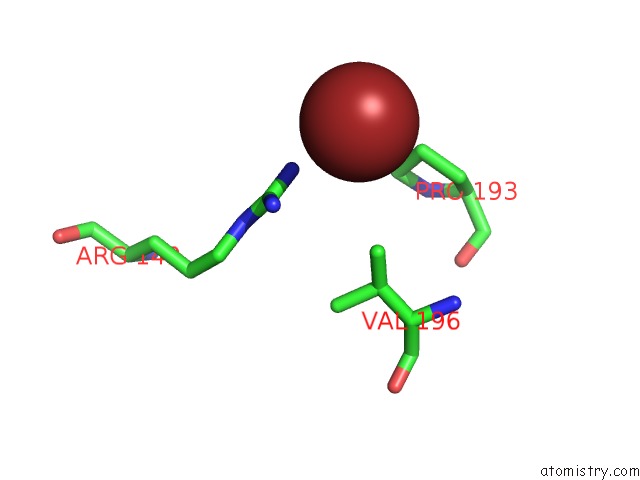
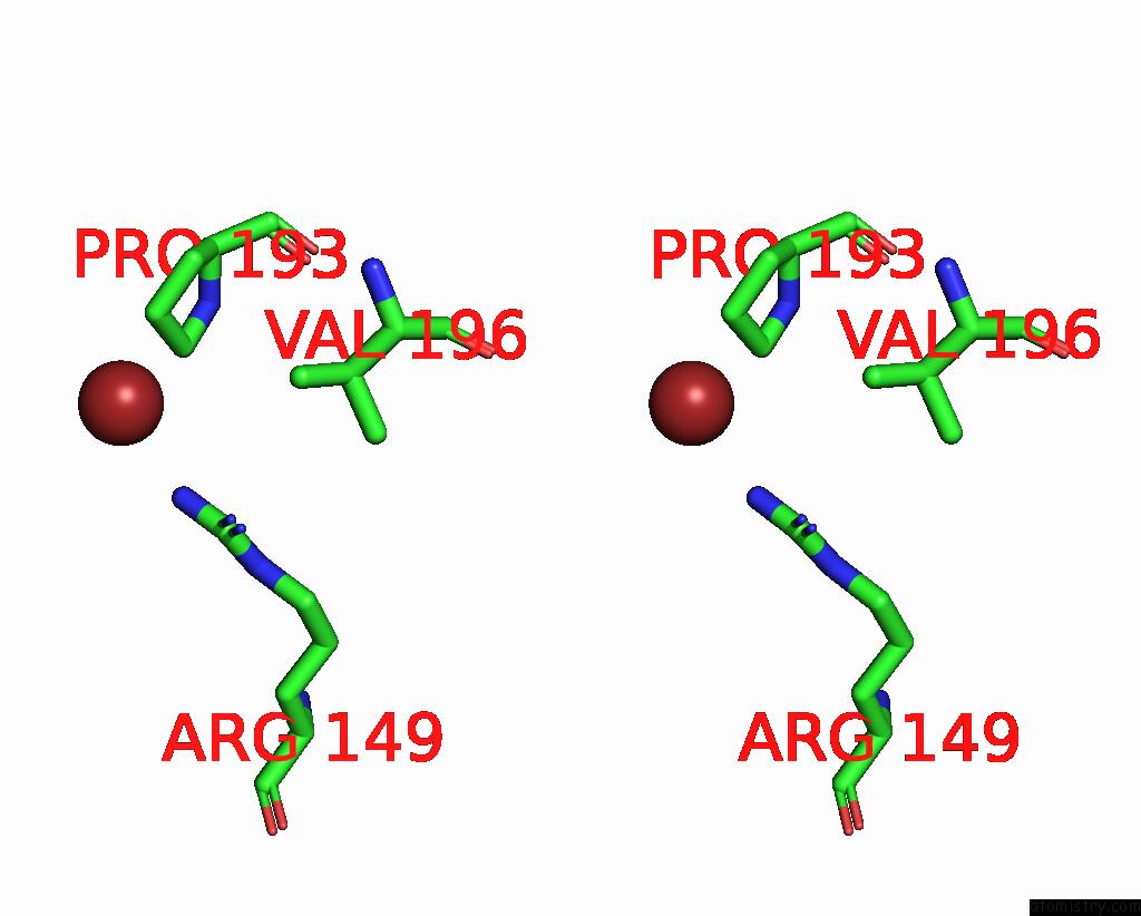
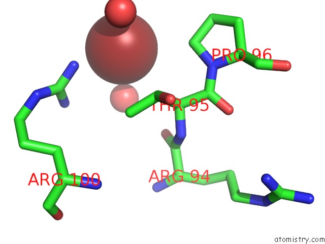
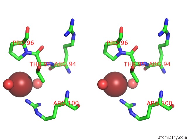
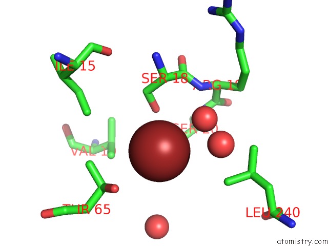
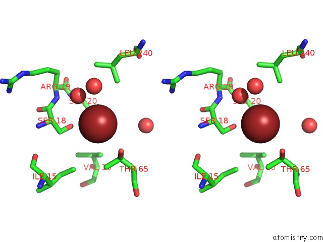
Bromine binding site 1 out of 7 in 4p4g
Go back to
Bromine binding site 1 out
of 7 in the Crystal Structure of Mycobacterium Tuberculosis Shikimate Dehydrogenase
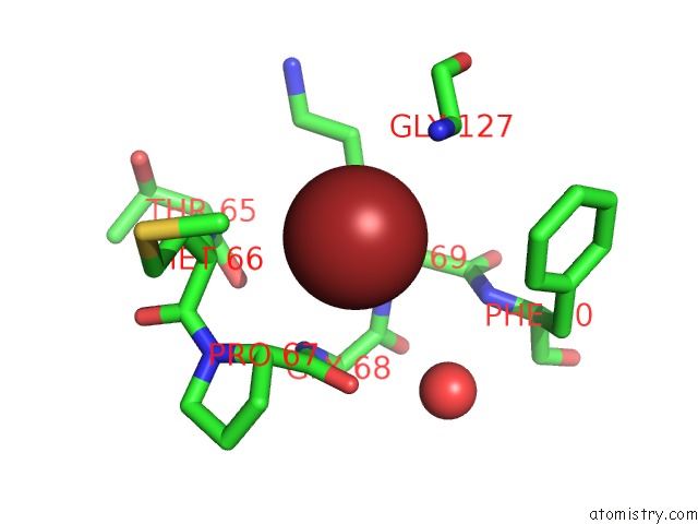
Mono view
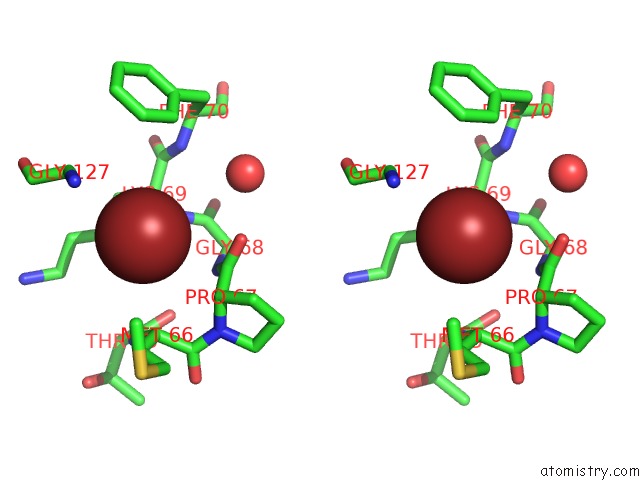
Stereo pair view
Mono view
Stereo pair view
A full contact list of Bromine with other atoms in the Br binding
site number 1 of Crystal Structure of Mycobacterium Tuberculosis Shikimate Dehydrogenase within 5.0Å range:
|
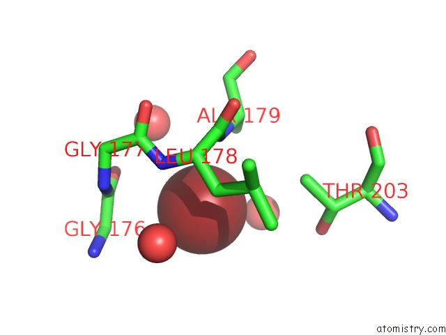
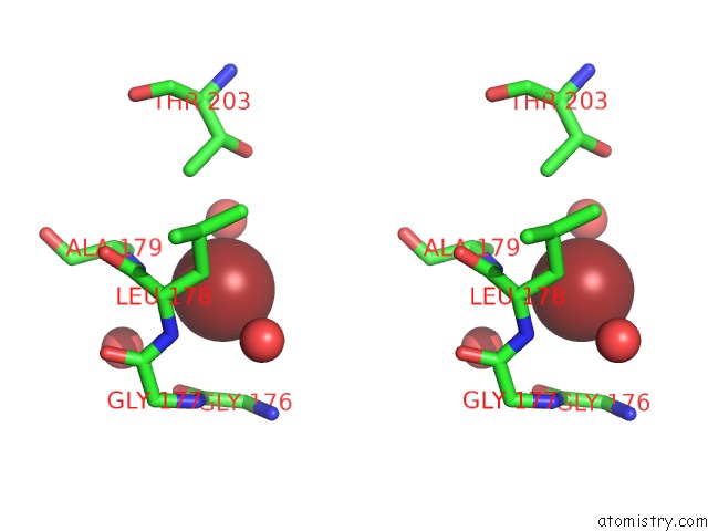
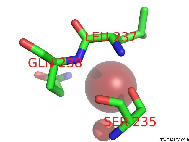
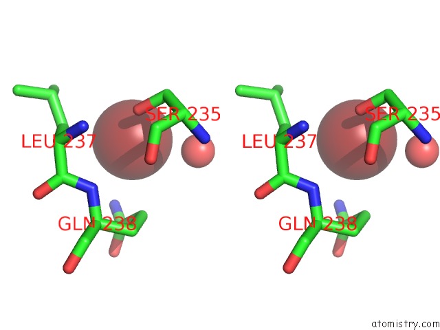
Bromine binding site 2 out of 7 in 4p4g
Go back to
Bromine binding site 2 out
of 7 in the Crystal Structure of Mycobacterium Tuberculosis Shikimate Dehydrogenase
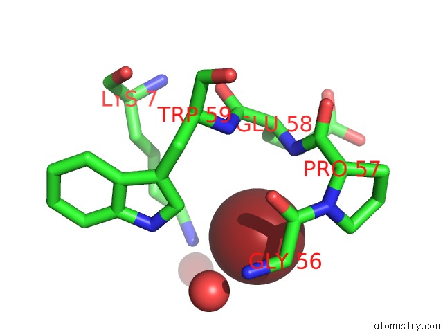
Mono view
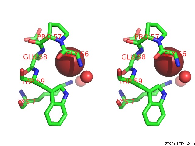
Stereo pair view
Mono view
Stereo pair view
A full contact list of Bromine with other atoms in the Br binding
site number 2 of Crystal Structure of Mycobacterium Tuberculosis Shikimate Dehydrogenase within 5.0Å range:
|
Bromine binding site 3 out of 7 in 4p4g
Go back to
Bromine binding site 3 out
of 7 in the Crystal Structure of Mycobacterium Tuberculosis Shikimate Dehydrogenase
Mono view
Stereo pair view
Mono view
Stereo pair view
A full contact list of Bromine with other atoms in the Br binding
site number 3 of Crystal Structure of Mycobacterium Tuberculosis Shikimate Dehydrogenase within 5.0Å range:
|
Bromine binding site 4 out of 7 in 4p4g
Go back to
Bromine binding site 4 out
of 7 in the Crystal Structure of Mycobacterium Tuberculosis Shikimate Dehydrogenase
Mono view
Stereo pair view
Mono view
Stereo pair view
A full contact list of Bromine with other atoms in the Br binding
site number 4 of Crystal Structure of Mycobacterium Tuberculosis Shikimate Dehydrogenase within 5.0Å range:
|
Bromine binding site 5 out of 7 in 4p4g
Go back to
Bromine binding site 5 out
of 7 in the Crystal Structure of Mycobacterium Tuberculosis Shikimate Dehydrogenase
Mono view
Stereo pair view
Mono view
Stereo pair view
A full contact list of Bromine with other atoms in the Br binding
site number 5 of Crystal Structure of Mycobacterium Tuberculosis Shikimate Dehydrogenase within 5.0Å range:
|
Bromine binding site 6 out of 7 in 4p4g
Go back to
Bromine binding site 6 out
of 7 in the Crystal Structure of Mycobacterium Tuberculosis Shikimate Dehydrogenase
Mono view
Stereo pair view
Mono view
Stereo pair view
A full contact list of Bromine with other atoms in the Br binding
site number 6 of Crystal Structure of Mycobacterium Tuberculosis Shikimate Dehydrogenase within 5.0Å range:
|
Bromine binding site 7 out of 7 in 4p4g
Go back to
Bromine binding site 7 out
of 7 in the Crystal Structure of Mycobacterium Tuberculosis Shikimate Dehydrogenase
Mono view
Stereo pair view
Mono view
Stereo pair view
A full contact list of Bromine with other atoms in the Br binding
site number 7 of Crystal Structure of Mycobacterium Tuberculosis Shikimate Dehydrogenase within 5.0Å range:
|
Reference:
M.Lalgondar,
J.C.Sacchettini.
Crystal Structure of Mycobacterium Tuberculosis Shikimate Dehydrogenase To Be Published.
Page generated: Mon Jul 7 07:13:59 2025
Last articles
Br in 7EJ8Br in 7D6J
Br in 7EFX
Br in 7EDT
Br in 7ECJ
Br in 7E0U
Br in 7ECD
Br in 7EBH
Br in 7E0S
Br in 7E0T