Bromine »
PDB 5cc3-5ev2 »
5ea4 »
Bromine in PDB 5ea4: Crystal Structure of Inhibitor Jnj-49153390 in Complex with Prefusion Rsv F Glycoprotein
Protein crystallography data
The structure of Crystal Structure of Inhibitor Jnj-49153390 in Complex with Prefusion Rsv F Glycoprotein, PDB code: 5ea4
was solved by
M.B.Battles,
J.S.Mclellan,
E.Arnoult,
D.Roymans,
J.P.Langedijk,
with X-Ray Crystallography technique. A brief refinement statistics is given in the table below:
| Resolution Low / High (Å) | 48.60 / 2.30 |
| Space group | P 41 3 2 |
| Cell size a, b, c (Å), α, β, γ (°) | 168.370, 168.370, 168.370, 90.00, 90.00, 90.00 |
| R / Rfree (%) | 17.9 / 21.4 |
Bromine Binding Sites:
The binding sites of Bromine atom in the Crystal Structure of Inhibitor Jnj-49153390 in Complex with Prefusion Rsv F Glycoprotein
(pdb code 5ea4). This binding sites where shown within
5.0 Angstroms radius around Bromine atom.
In total only one binding site of Bromine was determined in the Crystal Structure of Inhibitor Jnj-49153390 in Complex with Prefusion Rsv F Glycoprotein, PDB code: 5ea4:
In total only one binding site of Bromine was determined in the Crystal Structure of Inhibitor Jnj-49153390 in Complex with Prefusion Rsv F Glycoprotein, PDB code: 5ea4:
Bromine binding site 1 out of 1 in 5ea4
Go back to
Bromine binding site 1 out
of 1 in the Crystal Structure of Inhibitor Jnj-49153390 in Complex with Prefusion Rsv F Glycoprotein
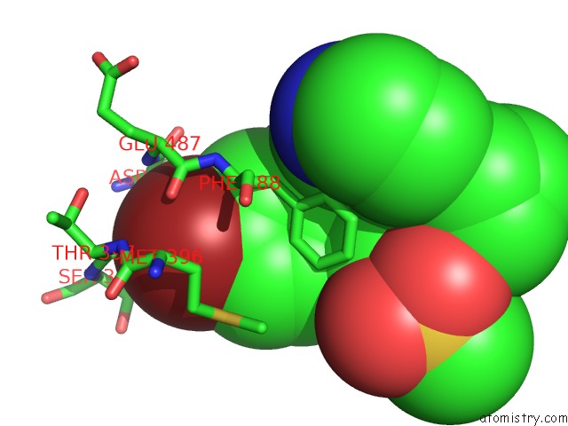
Mono view
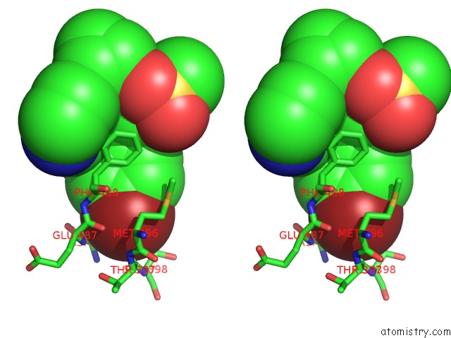
Stereo pair view
Mono view
Stereo pair view
A full contact list of Bromine with other atoms in the Br binding
site number 1 of Crystal Structure of Inhibitor Jnj-49153390 in Complex with Prefusion Rsv F Glycoprotein within 5.0Å range:
|
Reference:
M.B.Battles,
J.P.Langedijk,
P.Furmanova-Hollenstein,
S.Chaiwatpongsakorn,
H.M.Costello,
L.Kwanten,
L.Vranckx,
P.Vink,
S.Jaensch,
T.H.Jonckers,
A.Koul,
E.Arnoult,
M.E.Peeples,
D.Roymans,
J.S.Mclellan.
Molecular Mechanism of Respiratory Syncytial Virus Fusion Inhibitors. Nat.Chem.Biol. V. 12 87 2016.
ISSN: ESSN 1552-4469
PubMed: 26641933
DOI: 10.1038/NCHEMBIO.1982
Page generated: Wed Jul 10 23:28:03 2024
ISSN: ESSN 1552-4469
PubMed: 26641933
DOI: 10.1038/NCHEMBIO.1982
Last articles
Zn in 9J0NZn in 9J0O
Zn in 9J0P
Zn in 9FJX
Zn in 9EKB
Zn in 9C0F
Zn in 9CAH
Zn in 9CH0
Zn in 9CH3
Zn in 9CH1