Bromine »
PDB 5i74-5l3j »
5ii0 »
Bromine in PDB 5ii0: Crystal Structure of the Human Calcitonin Receptor Ectodomain in Complex with A Truncated Salmon Calcitonin Analogue
Protein crystallography data
The structure of Crystal Structure of the Human Calcitonin Receptor Ectodomain in Complex with A Truncated Salmon Calcitonin Analogue, PDB code: 5ii0
was solved by
E.Johansson,
S.Reedtz-Runge,
with X-Ray Crystallography technique. A brief refinement statistics is given in the table below:
| Resolution Low / High (Å) | 24.84 / 2.10 |
| Space group | C 1 2 1 |
| Cell size a, b, c (Å), α, β, γ (°) | 96.558, 113.165, 55.424, 90.00, 114.79, 90.00 |
| R / Rfree (%) | 17 / 20.6 |
Other elements in 5ii0:
The structure of Crystal Structure of the Human Calcitonin Receptor Ectodomain in Complex with A Truncated Salmon Calcitonin Analogue also contains other interesting chemical elements:
| Sodium | (Na) | 1 atom |
Bromine Binding Sites:
The binding sites of Bromine atom in the Crystal Structure of the Human Calcitonin Receptor Ectodomain in Complex with A Truncated Salmon Calcitonin Analogue
(pdb code 5ii0). This binding sites where shown within
5.0 Angstroms radius around Bromine atom.
In total 2 binding sites of Bromine where determined in the Crystal Structure of the Human Calcitonin Receptor Ectodomain in Complex with A Truncated Salmon Calcitonin Analogue, PDB code: 5ii0:
Jump to Bromine binding site number: 1; 2;
In total 2 binding sites of Bromine where determined in the Crystal Structure of the Human Calcitonin Receptor Ectodomain in Complex with A Truncated Salmon Calcitonin Analogue, PDB code: 5ii0:
Jump to Bromine binding site number: 1; 2;
Bromine binding site 1 out of 2 in 5ii0
Go back to
Bromine binding site 1 out
of 2 in the Crystal Structure of the Human Calcitonin Receptor Ectodomain in Complex with A Truncated Salmon Calcitonin Analogue
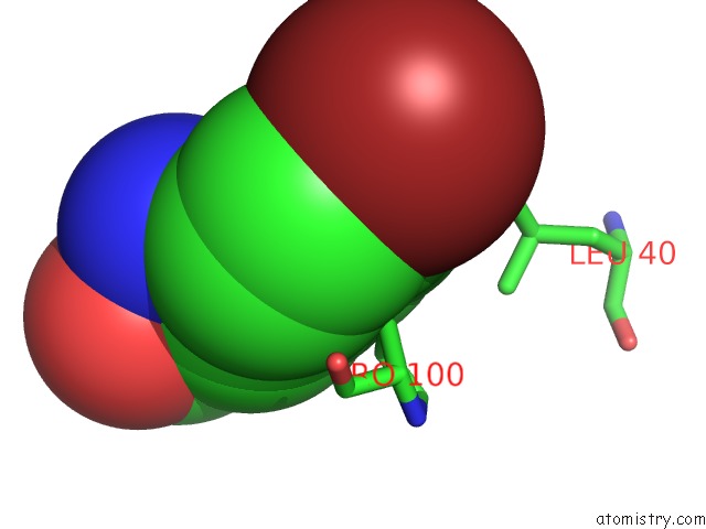
Mono view
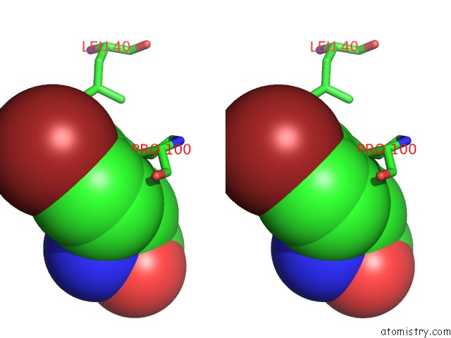
Stereo pair view
Mono view
Stereo pair view
A full contact list of Bromine with other atoms in the Br binding
site number 1 of Crystal Structure of the Human Calcitonin Receptor Ectodomain in Complex with A Truncated Salmon Calcitonin Analogue within 5.0Å range:
|
Bromine binding site 2 out of 2 in 5ii0
Go back to
Bromine binding site 2 out
of 2 in the Crystal Structure of the Human Calcitonin Receptor Ectodomain in Complex with A Truncated Salmon Calcitonin Analogue
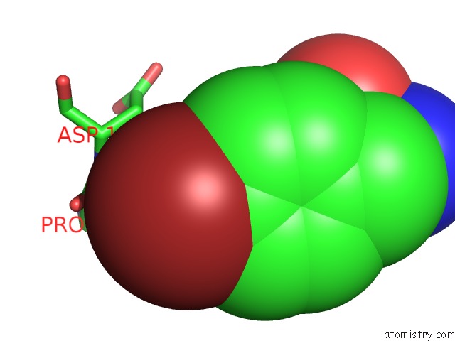
Mono view
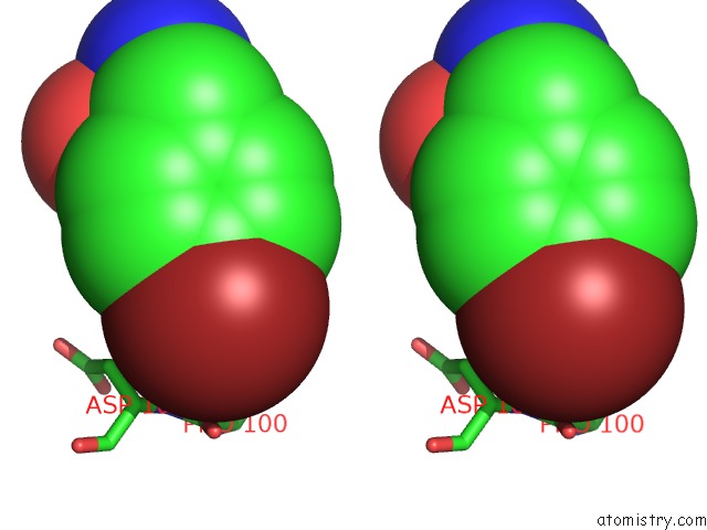
Stereo pair view
Mono view
Stereo pair view
A full contact list of Bromine with other atoms in the Br binding
site number 2 of Crystal Structure of the Human Calcitonin Receptor Ectodomain in Complex with A Truncated Salmon Calcitonin Analogue within 5.0Å range:
|
Reference:
E.Johansson,
J.L.Hansen,
A.M.Hansen,
A.C.Shaw,
P.Becker,
L.Schaffer,
S.Reedtz-Runge.
Type II Turn of Receptor-Bound Salmon Calcitonin Revealed By X-Ray Crystallography. J.Biol.Chem. V. 291 13689 2016.
ISSN: ESSN 1083-351X
PubMed: 27189946
DOI: 10.1074/JBC.M116.726034
Page generated: Thu Jul 11 00:04:30 2024
ISSN: ESSN 1083-351X
PubMed: 27189946
DOI: 10.1074/JBC.M116.726034
Last articles
Zn in 9J0NZn in 9J0O
Zn in 9J0P
Zn in 9FJX
Zn in 9EKB
Zn in 9C0F
Zn in 9CAH
Zn in 9CH0
Zn in 9CH3
Zn in 9CH1