Bromine »
PDB 5mrm-5o0j »
5ndh »
Bromine in PDB 5ndh: The Structure of the G. Violaceus Guanidine II Riboswitch P2 Stem-Loop
Protein crystallography data
The structure of The Structure of the G. Violaceus Guanidine II Riboswitch P2 Stem-Loop, PDB code: 5ndh
was solved by
L.Huang,
J.Wang,
D.M.J.Lilley,
with X-Ray Crystallography technique. A brief refinement statistics is given in the table below:
| Resolution Low / High (Å) | 50.17 / 1.81 |
| Space group | P 21 21 21 |
| Cell size a, b, c (Å), α, β, γ (°) | 48.727, 57.825, 100.862, 90.00, 90.00, 90.00 |
| R / Rfree (%) | 20.7 / 22.2 |
Other elements in 5ndh:
The structure of The Structure of the G. Violaceus Guanidine II Riboswitch P2 Stem-Loop also contains other interesting chemical elements:
| Magnesium | (Mg) | 1 atom |
| Sodium | (Na) | 4 atoms |
Bromine Binding Sites:
The binding sites of Bromine atom in the The Structure of the G. Violaceus Guanidine II Riboswitch P2 Stem-Loop
(pdb code 5ndh). This binding sites where shown within
5.0 Angstroms radius around Bromine atom.
In total 4 binding sites of Bromine where determined in the The Structure of the G. Violaceus Guanidine II Riboswitch P2 Stem-Loop, PDB code: 5ndh:
Jump to Bromine binding site number: 1; 2; 3; 4;
In total 4 binding sites of Bromine where determined in the The Structure of the G. Violaceus Guanidine II Riboswitch P2 Stem-Loop, PDB code: 5ndh:
Jump to Bromine binding site number: 1; 2; 3; 4;
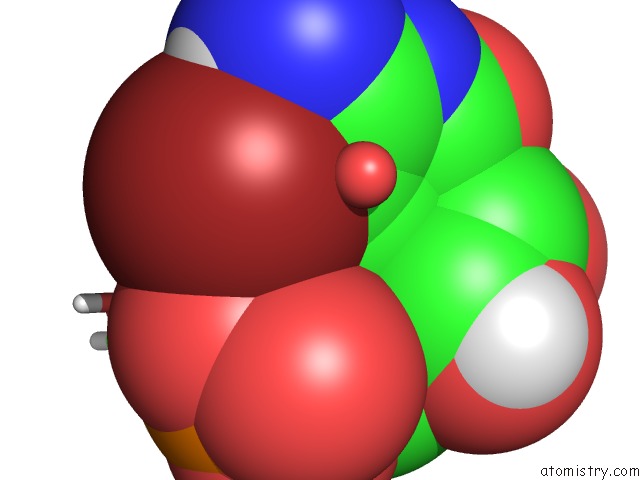
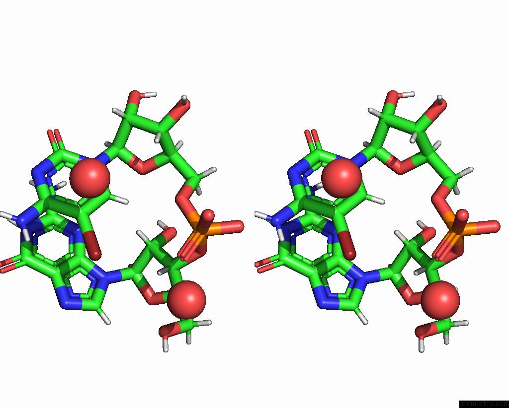
Bromine binding site 1 out of 4 in 5ndh
Go back to
Bromine binding site 1 out
of 4 in the The Structure of the G. Violaceus Guanidine II Riboswitch P2 Stem-Loop
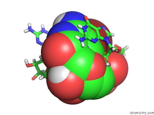
Mono view
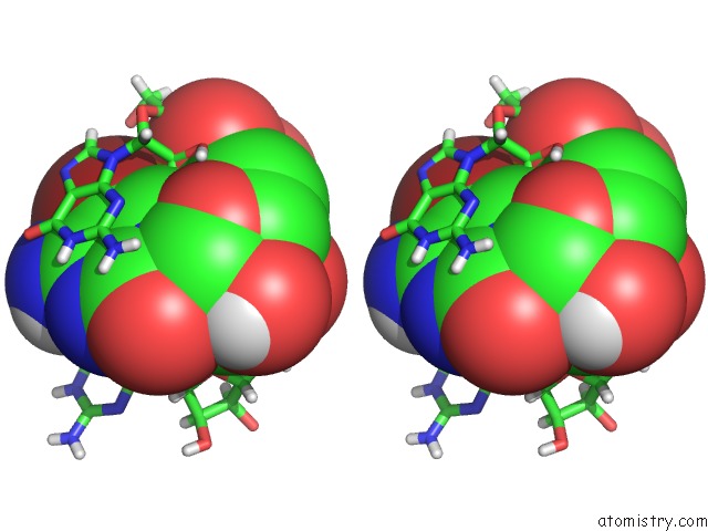
Stereo pair view
Mono view
Stereo pair view
A full contact list of Bromine with other atoms in the Br binding
site number 1 of The Structure of the G. Violaceus Guanidine II Riboswitch P2 Stem-Loop within 5.0Å range:
|
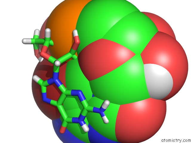
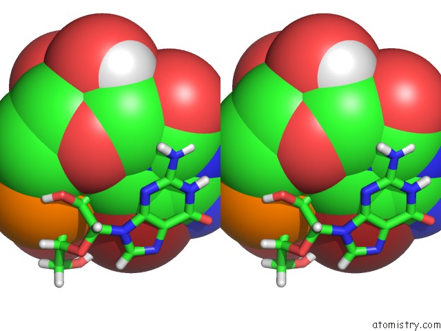
Bromine binding site 2 out of 4 in 5ndh
Go back to
Bromine binding site 2 out
of 4 in the The Structure of the G. Violaceus Guanidine II Riboswitch P2 Stem-Loop
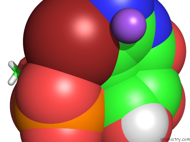
Mono view
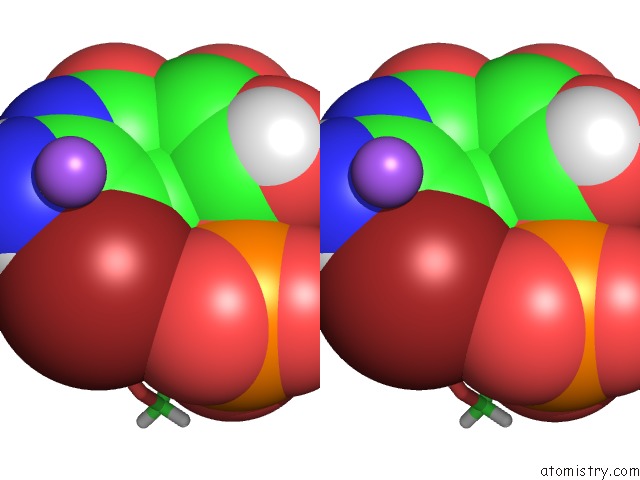
Stereo pair view
Mono view
Stereo pair view
A full contact list of Bromine with other atoms in the Br binding
site number 2 of The Structure of the G. Violaceus Guanidine II Riboswitch P2 Stem-Loop within 5.0Å range:
|
Bromine binding site 3 out of 4 in 5ndh
Go back to
Bromine binding site 3 out
of 4 in the The Structure of the G. Violaceus Guanidine II Riboswitch P2 Stem-Loop
Mono view
Stereo pair view
Mono view
Stereo pair view
A full contact list of Bromine with other atoms in the Br binding
site number 3 of The Structure of the G. Violaceus Guanidine II Riboswitch P2 Stem-Loop within 5.0Å range:
|
Bromine binding site 4 out of 4 in 5ndh
Go back to
Bromine binding site 4 out
of 4 in the The Structure of the G. Violaceus Guanidine II Riboswitch P2 Stem-Loop
Mono view
Stereo pair view
Mono view
Stereo pair view
A full contact list of Bromine with other atoms in the Br binding
site number 4 of The Structure of the G. Violaceus Guanidine II Riboswitch P2 Stem-Loop within 5.0Å range:
|
Reference:
L.Huang,
J.Wang,
D.M.J.Lilley.
The Structure of the Guanidine-II Riboswitch. Cell Chem Biol V. 24 695 2017.
ISSN: ESSN 2451-9456
PubMed: 28529131
DOI: 10.1016/J.CHEMBIOL.2017.05.014
Page generated: Thu Jul 11 00:27:40 2024
ISSN: ESSN 2451-9456
PubMed: 28529131
DOI: 10.1016/J.CHEMBIOL.2017.05.014
Last articles
Zn in 9MJ5Zn in 9HNW
Zn in 9G0L
Zn in 9FNE
Zn in 9DZN
Zn in 9E0I
Zn in 9D32
Zn in 9DAK
Zn in 8ZXC
Zn in 8ZUF