Bromine »
PDB 5y94-6c2x »
6arz »
Bromine in PDB 6arz: Structure of A Phage Anti-Crispr Protein
Protein crystallography data
The structure of Structure of A Phage Anti-Crispr Protein, PDB code: 6arz
was solved by
C.Calmettes,
M.Shah,
A.Pawluk,
A.R.Davidson,
K.L.Maxwell,
T.F.Moraes,
with X-Ray Crystallography technique. A brief refinement statistics is given in the table below:
| Resolution Low / High (Å) | 48.18 / 2.50 |
| Space group | C 1 2 1 |
| Cell size a, b, c (Å), α, β, γ (°) | 98.137, 63.959, 59.701, 90.00, 100.94, 90.00 |
| R / Rfree (%) | 18 / 22.1 |
Bromine Binding Sites:
The binding sites of Bromine atom in the Structure of A Phage Anti-Crispr Protein
(pdb code 6arz). This binding sites where shown within
5.0 Angstroms radius around Bromine atom.
In total 5 binding sites of Bromine where determined in the Structure of A Phage Anti-Crispr Protein, PDB code: 6arz:
Jump to Bromine binding site number: 1; 2; 3; 4; 5;
In total 5 binding sites of Bromine where determined in the Structure of A Phage Anti-Crispr Protein, PDB code: 6arz:
Jump to Bromine binding site number: 1; 2; 3; 4; 5;
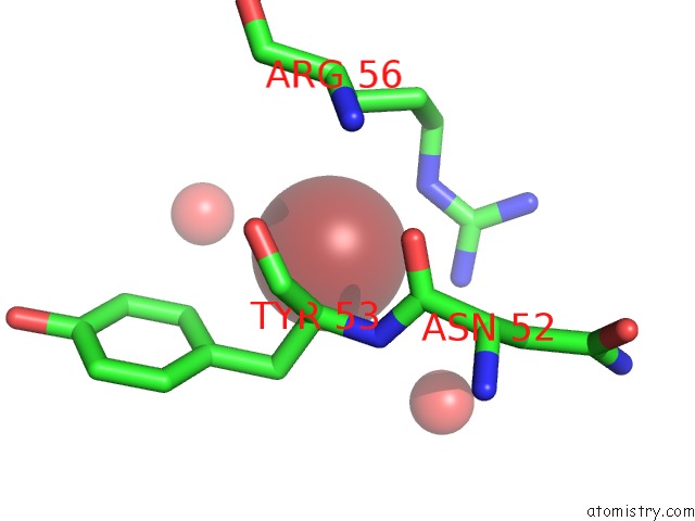
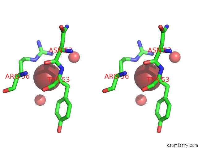
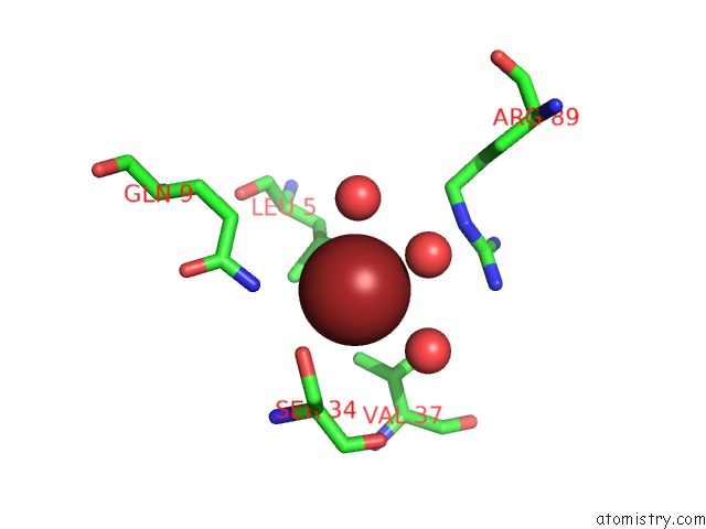
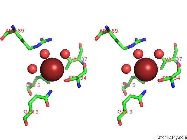
Bromine binding site 1 out of 5 in 6arz
Go back to
Bromine binding site 1 out
of 5 in the Structure of A Phage Anti-Crispr Protein
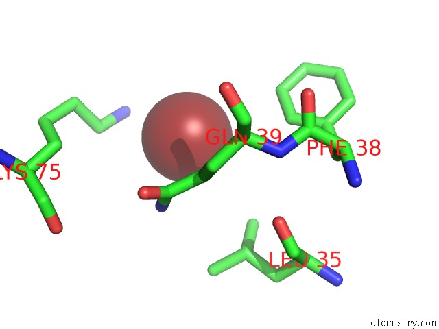
Mono view
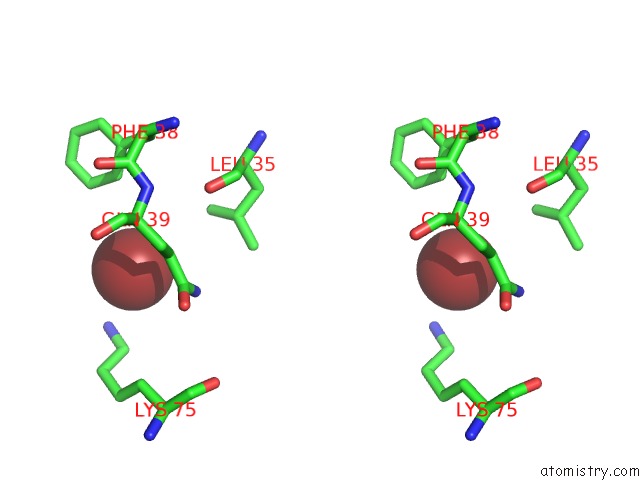
Stereo pair view
Mono view
Stereo pair view
A full contact list of Bromine with other atoms in the Br binding
site number 1 of Structure of A Phage Anti-Crispr Protein within 5.0Å range:
|
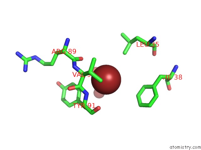
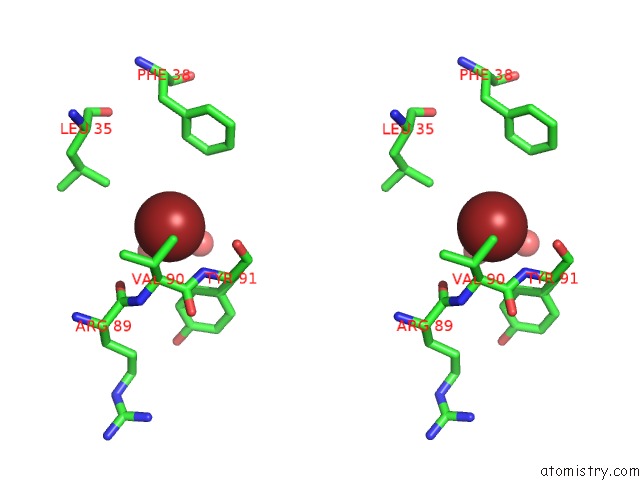
Bromine binding site 2 out of 5 in 6arz
Go back to
Bromine binding site 2 out
of 5 in the Structure of A Phage Anti-Crispr Protein
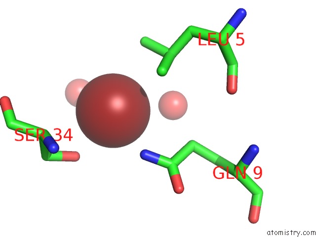
Mono view
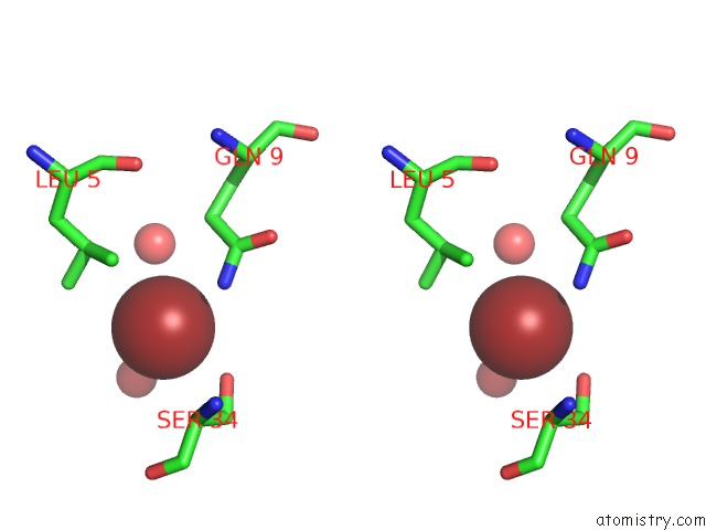
Stereo pair view
Mono view
Stereo pair view
A full contact list of Bromine with other atoms in the Br binding
site number 2 of Structure of A Phage Anti-Crispr Protein within 5.0Å range:
|
Bromine binding site 3 out of 5 in 6arz
Go back to
Bromine binding site 3 out
of 5 in the Structure of A Phage Anti-Crispr Protein
Mono view
Stereo pair view
Mono view
Stereo pair view
A full contact list of Bromine with other atoms in the Br binding
site number 3 of Structure of A Phage Anti-Crispr Protein within 5.0Å range:
|
Bromine binding site 4 out of 5 in 6arz
Go back to
Bromine binding site 4 out
of 5 in the Structure of A Phage Anti-Crispr Protein
Mono view
Stereo pair view
Mono view
Stereo pair view
A full contact list of Bromine with other atoms in the Br binding
site number 4 of Structure of A Phage Anti-Crispr Protein within 5.0Å range:
|
Bromine binding site 5 out of 5 in 6arz
Go back to
Bromine binding site 5 out
of 5 in the Structure of A Phage Anti-Crispr Protein
Mono view
Stereo pair view
Mono view
Stereo pair view
A full contact list of Bromine with other atoms in the Br binding
site number 5 of Structure of A Phage Anti-Crispr Protein within 5.0Å range:
|
Reference:
A.Pawluk,
M.Shah,
M.Mejdani,
C.Calmettes,
T.F.Moraes,
A.R.Davidson,
K.L.Maxwell.
Disabling A Type I-E Crispr-Cas Nuclease with A Bacteriophage-Encoded Anti-Crispr Protein. Mbio V. 8 2017.
ISSN: ESSN 2150-7511
PubMed: 29233895
DOI: 10.1128/MBIO.01751-17
Page generated: Thu Jul 11 01:28:07 2024
ISSN: ESSN 2150-7511
PubMed: 29233895
DOI: 10.1128/MBIO.01751-17
Last articles
Zn in 9MJ5Zn in 9HNW
Zn in 9G0L
Zn in 9FNE
Zn in 9DZN
Zn in 9E0I
Zn in 9D32
Zn in 9DAK
Zn in 8ZXC
Zn in 8ZUF