Bromine »
PDB 6hbt-6lg9 »
6iyx »
Bromine in PDB 6iyx: Crystal Structure Analysis of A Eukaryotic Membrane Protein
Protein crystallography data
The structure of Crystal Structure Analysis of A Eukaryotic Membrane Protein, PDB code: 6iyx
was solved by
Y.Zeng,
X.H.Wang,
F.Gao,
M.Su,
Y.H.Chen,
with X-Ray Crystallography technique. A brief refinement statistics is given in the table below:
| Resolution Low / High (Å) | 46.27 / 1.80 |
| Space group | P 63 2 2 |
| Cell size a, b, c (Å), α, β, γ (°) | 126.723, 126.723, 102.041, 90.00, 90.00, 120.00 |
| R / Rfree (%) | 20.2 / 21.2 |
Other elements in 6iyx:
The structure of Crystal Structure Analysis of A Eukaryotic Membrane Protein also contains other interesting chemical elements:
| Calcium | (Ca) | 1 atom |
Bromine Binding Sites:
The binding sites of Bromine atom in the Crystal Structure Analysis of A Eukaryotic Membrane Protein
(pdb code 6iyx). This binding sites where shown within
5.0 Angstroms radius around Bromine atom.
In total 6 binding sites of Bromine where determined in the Crystal Structure Analysis of A Eukaryotic Membrane Protein, PDB code: 6iyx:
Jump to Bromine binding site number: 1; 2; 3; 4; 5; 6;
In total 6 binding sites of Bromine where determined in the Crystal Structure Analysis of A Eukaryotic Membrane Protein, PDB code: 6iyx:
Jump to Bromine binding site number: 1; 2; 3; 4; 5; 6;
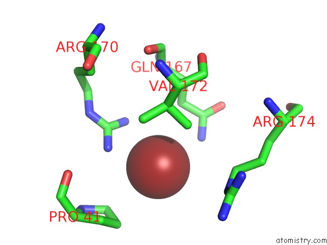
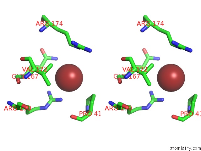
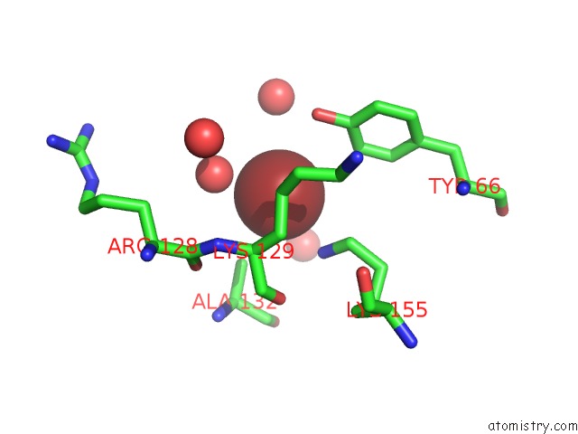
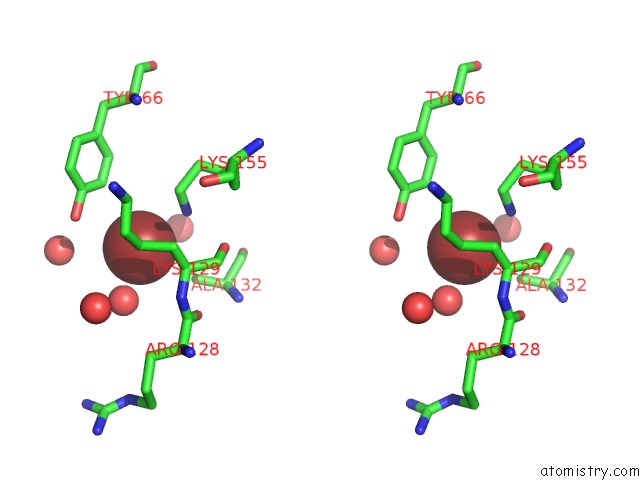
Bromine binding site 1 out of 6 in 6iyx
Go back to
Bromine binding site 1 out
of 6 in the Crystal Structure Analysis of A Eukaryotic Membrane Protein
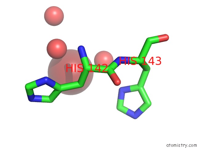
Mono view
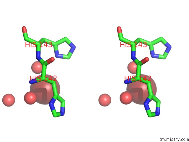
Stereo pair view
Mono view
Stereo pair view
A full contact list of Bromine with other atoms in the Br binding
site number 1 of Crystal Structure Analysis of A Eukaryotic Membrane Protein within 5.0Å range:
|
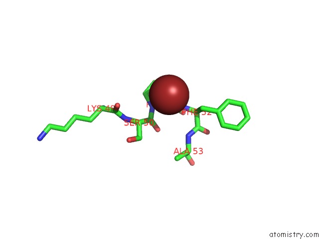
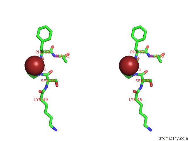
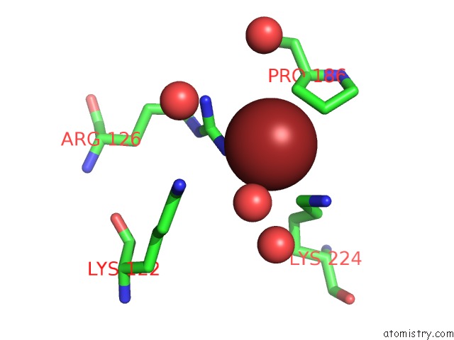
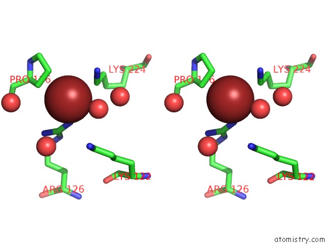
Bromine binding site 2 out of 6 in 6iyx
Go back to
Bromine binding site 2 out
of 6 in the Crystal Structure Analysis of A Eukaryotic Membrane Protein
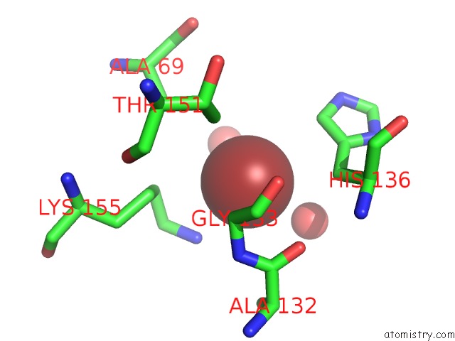
Mono view
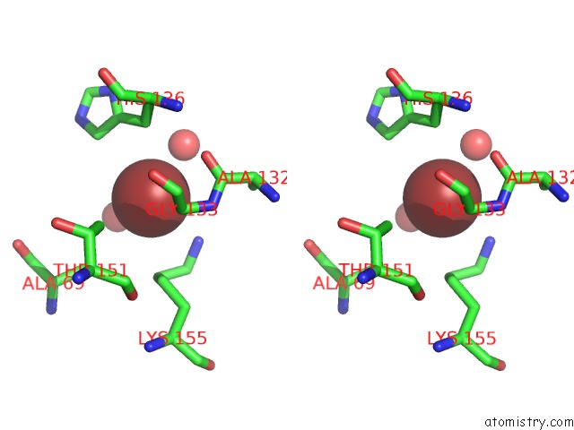
Stereo pair view
Mono view
Stereo pair view
A full contact list of Bromine with other atoms in the Br binding
site number 2 of Crystal Structure Analysis of A Eukaryotic Membrane Protein within 5.0Å range:
|
Bromine binding site 3 out of 6 in 6iyx
Go back to
Bromine binding site 3 out
of 6 in the Crystal Structure Analysis of A Eukaryotic Membrane Protein
Mono view
Stereo pair view
Mono view
Stereo pair view
A full contact list of Bromine with other atoms in the Br binding
site number 3 of Crystal Structure Analysis of A Eukaryotic Membrane Protein within 5.0Å range:
|
Bromine binding site 4 out of 6 in 6iyx
Go back to
Bromine binding site 4 out
of 6 in the Crystal Structure Analysis of A Eukaryotic Membrane Protein
Mono view
Stereo pair view
Mono view
Stereo pair view
A full contact list of Bromine with other atoms in the Br binding
site number 4 of Crystal Structure Analysis of A Eukaryotic Membrane Protein within 5.0Å range:
|
Bromine binding site 5 out of 6 in 6iyx
Go back to
Bromine binding site 5 out
of 6 in the Crystal Structure Analysis of A Eukaryotic Membrane Protein
Mono view
Stereo pair view
Mono view
Stereo pair view
A full contact list of Bromine with other atoms in the Br binding
site number 5 of Crystal Structure Analysis of A Eukaryotic Membrane Protein within 5.0Å range:
|
Bromine binding site 6 out of 6 in 6iyx
Go back to
Bromine binding site 6 out
of 6 in the Crystal Structure Analysis of A Eukaryotic Membrane Protein
Mono view
Stereo pair view
Mono view
Stereo pair view
A full contact list of Bromine with other atoms in the Br binding
site number 6 of Crystal Structure Analysis of A Eukaryotic Membrane Protein within 5.0Å range:
|
Reference:
X.H.Wang,
M.Su,
F.Gao,
W.Xie,
Y.Zeng,
D.L.Li,
X.L.Liu,
H.Zhao,
L.Qin,
F.Li,
Q.Liu,
O.B.Clarke,
S.M.Lam,
G.H.Shui,
W.A.Hendrickson,
Y.H.Chen.
Structural Basis For Activity of Tric Counter-Ion Channels in Calcium Release. Proc.Natl.Acad.Sci.Usa V. 116 4238 2019.
ISSN: ESSN 1091-6490
PubMed: 30770441
DOI: 10.1073/PNAS.1817271116
Page generated: Mon Jul 7 10:03:42 2025
ISSN: ESSN 1091-6490
PubMed: 30770441
DOI: 10.1073/PNAS.1817271116
Last articles
Fe in 2YXOFe in 2YRS
Fe in 2YXC
Fe in 2YNM
Fe in 2YVJ
Fe in 2YP1
Fe in 2YU2
Fe in 2YU1
Fe in 2YQB
Fe in 2YOO