Bromine »
PDB 6lue-6puu »
6m5b »
Bromine in PDB 6m5b: X-Ray Crystal Structure of Cyclic-Pip and Dna Complex in A Reverse Binding Orientation
Protein crystallography data
The structure of X-Ray Crystal Structure of Cyclic-Pip and Dna Complex in A Reverse Binding Orientation, PDB code: 6m5b
was solved by
K.Abe,
Y.Hirose,
H.Eki,
K.Takeda,
T.Bando,
M.Endo,
H.Sugiyama,
with X-Ray Crystallography technique. A brief refinement statistics is given in the table below:
| Resolution Low / High (Å) | 29.07 / 1.50 |
| Space group | I 41 3 2 |
| Cell size a, b, c (Å), α, β, γ (°) | 82.235, 82.235, 82.235, 90.00, 90.00, 90.00 |
| R / Rfree (%) | 18.4 / 18.9 |
Other elements in 6m5b:
The structure of X-Ray Crystal Structure of Cyclic-Pip and Dna Complex in A Reverse Binding Orientation also contains other interesting chemical elements:
| Magnesium | (Mg) | 1 atom |
| Sodium | (Na) | 1 atom |
Bromine Binding Sites:
The binding sites of Bromine atom in the X-Ray Crystal Structure of Cyclic-Pip and Dna Complex in A Reverse Binding Orientation
(pdb code 6m5b). This binding sites where shown within
5.0 Angstroms radius around Bromine atom.
In total only one binding site of Bromine was determined in the X-Ray Crystal Structure of Cyclic-Pip and Dna Complex in A Reverse Binding Orientation, PDB code: 6m5b:
In total only one binding site of Bromine was determined in the X-Ray Crystal Structure of Cyclic-Pip and Dna Complex in A Reverse Binding Orientation, PDB code: 6m5b:
Bromine binding site 1 out of 1 in 6m5b
Go back to
Bromine binding site 1 out
of 1 in the X-Ray Crystal Structure of Cyclic-Pip and Dna Complex in A Reverse Binding Orientation
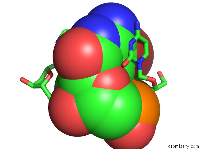
Mono view
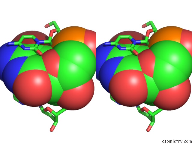
Stereo pair view
Mono view
Stereo pair view
A full contact list of Bromine with other atoms in the Br binding
site number 1 of X-Ray Crystal Structure of Cyclic-Pip and Dna Complex in A Reverse Binding Orientation within 5.0Å range:
|
Reference:
K.Abe,
Y.Hirose,
H.Eki,
K.Takeda,
T.Bando,
M.Endo,
H.Sugiyama.
X-Ray Crystal Structure of A Cyclic-Pip-Dna Complex in the Reverse-Binding Orientation. J.Am.Chem.Soc. V. 142 10544 2020.
ISSN: ESSN 1520-5126
PubMed: 32401492
DOI: 10.1021/JACS.0C03972
Page generated: Thu Jul 11 02:16:18 2024
ISSN: ESSN 1520-5126
PubMed: 32401492
DOI: 10.1021/JACS.0C03972
Last articles
Zn in 9J0NZn in 9J0O
Zn in 9J0P
Zn in 9FJX
Zn in 9EKB
Zn in 9C0F
Zn in 9CAH
Zn in 9CH0
Zn in 9CH3
Zn in 9CH1