Bromine »
PDB 6pv8-6ror »
6qn3 »
Bromine in PDB 6qn3: Structure of the Glutamine II Riboswitch
Protein crystallography data
The structure of Structure of the Glutamine II Riboswitch, PDB code: 6qn3
was solved by
L.Huang,
D.M.J.Lilley,
with X-Ray Crystallography technique. A brief refinement statistics is given in the table below:
| Resolution Low / High (Å) | 47.00 / 2.30 |
| Space group | C 2 2 21 |
| Cell size a, b, c (Å), α, β, γ (°) | 76.502, 115.725, 80.582, 90.00, 90.00, 90.00 |
| R / Rfree (%) | 24.2 / 29.4 |
Other elements in 6qn3:
The structure of Structure of the Glutamine II Riboswitch also contains other interesting chemical elements:
| Magnesium | (Mg) | 4 atoms |
Bromine Binding Sites:
The binding sites of Bromine atom in the Structure of the Glutamine II Riboswitch
(pdb code 6qn3). This binding sites where shown within
5.0 Angstroms radius around Bromine atom.
In total 4 binding sites of Bromine where determined in the Structure of the Glutamine II Riboswitch, PDB code: 6qn3:
Jump to Bromine binding site number: 1; 2; 3; 4;
In total 4 binding sites of Bromine where determined in the Structure of the Glutamine II Riboswitch, PDB code: 6qn3:
Jump to Bromine binding site number: 1; 2; 3; 4;
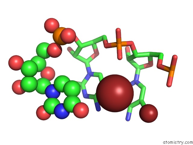
Bromine binding site 1 out of 4 in 6qn3
Go back to
Bromine binding site 1 out
of 4 in the Structure of the Glutamine II Riboswitch
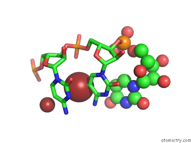
Mono view
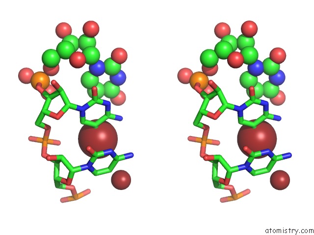
Stereo pair view
Mono view
Stereo pair view
A full contact list of Bromine with other atoms in the Br binding
site number 1 of Structure of the Glutamine II Riboswitch within 5.0Å range:
|
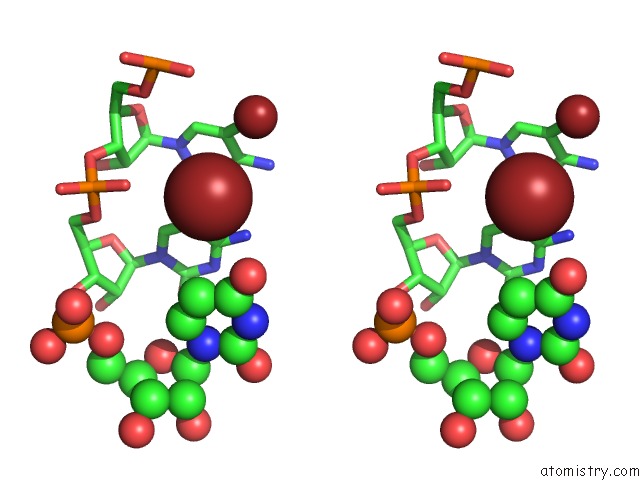
Bromine binding site 2 out of 4 in 6qn3
Go back to
Bromine binding site 2 out
of 4 in the Structure of the Glutamine II Riboswitch
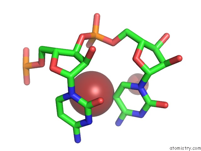
Mono view
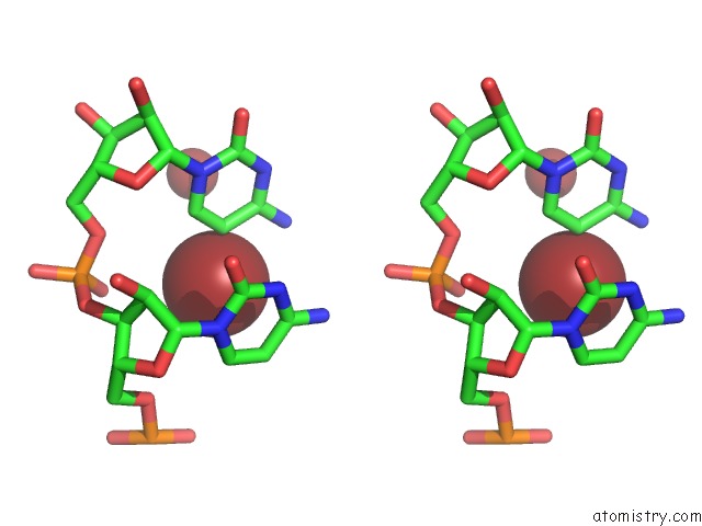
Stereo pair view
Mono view
Stereo pair view
A full contact list of Bromine with other atoms in the Br binding
site number 2 of Structure of the Glutamine II Riboswitch within 5.0Å range:
|
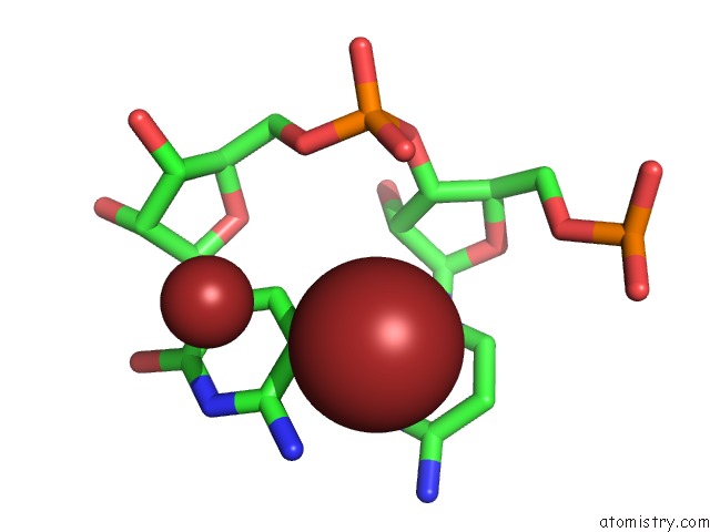
Bromine binding site 3 out of 4 in 6qn3
Go back to
Bromine binding site 3 out
of 4 in the Structure of the Glutamine II Riboswitch
Mono view
Stereo pair view
Mono view
Stereo pair view
A full contact list of Bromine with other atoms in the Br binding
site number 3 of Structure of the Glutamine II Riboswitch within 5.0Å range:
|
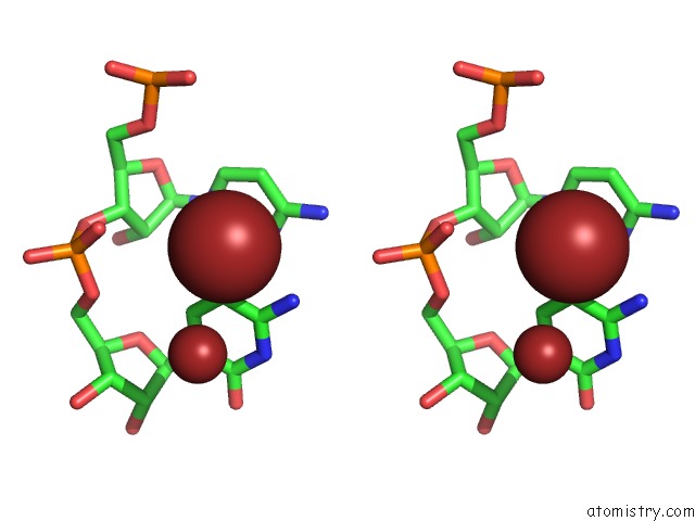
Bromine binding site 4 out of 4 in 6qn3
Go back to
Bromine binding site 4 out
of 4 in the Structure of the Glutamine II Riboswitch
Mono view
Stereo pair view
Mono view
Stereo pair view
A full contact list of Bromine with other atoms in the Br binding
site number 4 of Structure of the Glutamine II Riboswitch within 5.0Å range:
|
Reference:
L.Huang,
J.Wang,
A.M.Watkins,
R.Das,
D.M.J.Lilley.
Structure and Ligand Binding of the Glutamine-II Riboswitch. Nucleic Acids Res. V. 47 7666 2019.
ISSN: ESSN 1362-4962
PubMed: 31216023
DOI: 10.1093/NAR/GKZ539
Page generated: Mon Jul 7 10:17:30 2025
ISSN: ESSN 1362-4962
PubMed: 31216023
DOI: 10.1093/NAR/GKZ539
Last articles
Ca in 5L7PCa in 5L41
Ca in 5L3U
Ca in 5L73
Ca in 5L79
Ca in 5L30
Ca in 5L2Z
Ca in 5L1I
Ca in 5L2X
Ca in 5L2Y