Bromine »
PDB 6pv8-6ror »
6rbx »
Bromine in PDB 6rbx: Crystal Structure of Nad Kinase 1 From Listeria Monocytogenes in Complexe with An Adenine Derivative
Enzymatic activity of Crystal Structure of Nad Kinase 1 From Listeria Monocytogenes in Complexe with An Adenine Derivative
All present enzymatic activity of Crystal Structure of Nad Kinase 1 From Listeria Monocytogenes in Complexe with An Adenine Derivative:
2.7.1.23;
2.7.1.23;
Protein crystallography data
The structure of Crystal Structure of Nad Kinase 1 From Listeria Monocytogenes in Complexe with An Adenine Derivative, PDB code: 6rbx
was solved by
M.Gelin,
G.Labesse,
with X-Ray Crystallography technique. A brief refinement statistics is given in the table below:
| Resolution Low / High (Å) | 59.73 / 2.47 |
| Space group | I 2 2 2 |
| Cell size a, b, c (Å), α, β, γ (°) | 63.340, 75.710, 119.450, 90.00, 90.00, 90.00 |
| R / Rfree (%) | 21.4 / 25.9 |
Bromine Binding Sites:
The binding sites of Bromine atom in the Crystal Structure of Nad Kinase 1 From Listeria Monocytogenes in Complexe with An Adenine Derivative
(pdb code 6rbx). This binding sites where shown within
5.0 Angstroms radius around Bromine atom.
In total only one binding site of Bromine was determined in the Crystal Structure of Nad Kinase 1 From Listeria Monocytogenes in Complexe with An Adenine Derivative, PDB code: 6rbx:
In total only one binding site of Bromine was determined in the Crystal Structure of Nad Kinase 1 From Listeria Monocytogenes in Complexe with An Adenine Derivative, PDB code: 6rbx:
Bromine binding site 1 out of 1 in 6rbx
Go back to
Bromine binding site 1 out
of 1 in the Crystal Structure of Nad Kinase 1 From Listeria Monocytogenes in Complexe with An Adenine Derivative
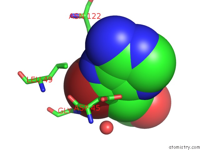
Mono view
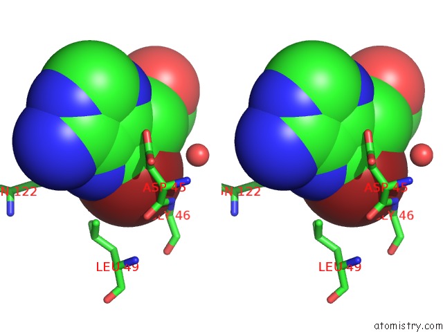
Stereo pair view
Mono view
Stereo pair view
A full contact list of Bromine with other atoms in the Br binding
site number 1 of Crystal Structure of Nad Kinase 1 From Listeria Monocytogenes in Complexe with An Adenine Derivative within 5.0Å range:
|
Reference:
M.Gelin,
J.Paoletti,
M.A.Nahori,
V.Huteau,
C.Leseigneur,
G.Jouvion,
L.Dugue,
D.Clement,
J.L.Pons,
L.Assairi,
S.Pochet,
G.Labesse,
O.Dussurget.
From Substrate to Fragments to Inhibitor Activein Vivoagainststaphylococcus Aureus. Acs Infect Dis. 2020.
ISSN: ESSN 2373-8227
PubMed: 32017533
DOI: 10.1021/ACSINFECDIS.9B00368
Page generated: Mon Jul 7 10:19:15 2025
ISSN: ESSN 2373-8227
PubMed: 32017533
DOI: 10.1021/ACSINFECDIS.9B00368
Last articles
Cl in 8CAACl in 8CAM
Cl in 8C9F
Cl in 8C9G
Cl in 8C9E
Cl in 8C8D
Cl in 8C8S
Cl in 8C8J
Cl in 8C7J
Cl in 8C7Y