Bromine »
PDB 6tu4-6vrk »
6tvn »
Bromine in PDB 6tvn: Crystal Structure of 5-Bromoindoline-2,3-Dione Covalently Bound to the pH Domain of Bruton'S Tyrosine Kinase
Enzymatic activity of Crystal Structure of 5-Bromoindoline-2,3-Dione Covalently Bound to the pH Domain of Bruton'S Tyrosine Kinase
All present enzymatic activity of Crystal Structure of 5-Bromoindoline-2,3-Dione Covalently Bound to the pH Domain of Bruton'S Tyrosine Kinase:
2.7.10.2;
2.7.10.2;
Protein crystallography data
The structure of Crystal Structure of 5-Bromoindoline-2,3-Dione Covalently Bound to the pH Domain of Bruton'S Tyrosine Kinase, PDB code: 6tvn
was solved by
P.Brear,
J.Wagstaff,
M.Hyvonen,
with X-Ray Crystallography technique. A brief refinement statistics is given in the table below:
| Resolution Low / High (Å) | 66.45 / 2.31 |
| Space group | P 1 21 1 |
| Cell size a, b, c (Å), α, β, γ (°) | 67.775, 65.400, 79.752, 90.00, 101.34, 90.00 |
| R / Rfree (%) | 22.5 / 25.7 |
Other elements in 6tvn:
The structure of Crystal Structure of 5-Bromoindoline-2,3-Dione Covalently Bound to the pH Domain of Bruton'S Tyrosine Kinase also contains other interesting chemical elements:
| Magnesium | (Mg) | 2 atoms |
| Zinc | (Zn) | 4 atoms |
Bromine Binding Sites:
The binding sites of Bromine atom in the Crystal Structure of 5-Bromoindoline-2,3-Dione Covalently Bound to the pH Domain of Bruton'S Tyrosine Kinase
(pdb code 6tvn). This binding sites where shown within
5.0 Angstroms radius around Bromine atom.
In total 4 binding sites of Bromine where determined in the Crystal Structure of 5-Bromoindoline-2,3-Dione Covalently Bound to the pH Domain of Bruton'S Tyrosine Kinase, PDB code: 6tvn:
Jump to Bromine binding site number: 1; 2; 3; 4;
In total 4 binding sites of Bromine where determined in the Crystal Structure of 5-Bromoindoline-2,3-Dione Covalently Bound to the pH Domain of Bruton'S Tyrosine Kinase, PDB code: 6tvn:
Jump to Bromine binding site number: 1; 2; 3; 4;
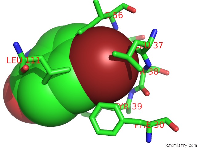
Bromine binding site 1 out of 4 in 6tvn
Go back to
Bromine binding site 1 out
of 4 in the Crystal Structure of 5-Bromoindoline-2,3-Dione Covalently Bound to the pH Domain of Bruton'S Tyrosine Kinase
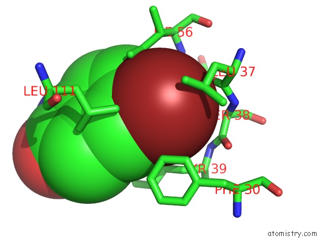
Mono view
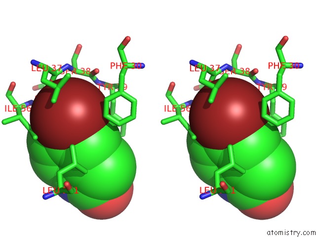
Stereo pair view
Mono view
Stereo pair view
A full contact list of Bromine with other atoms in the Br binding
site number 1 of Crystal Structure of 5-Bromoindoline-2,3-Dione Covalently Bound to the pH Domain of Bruton'S Tyrosine Kinase within 5.0Å range:
|
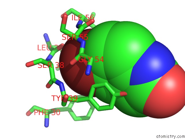
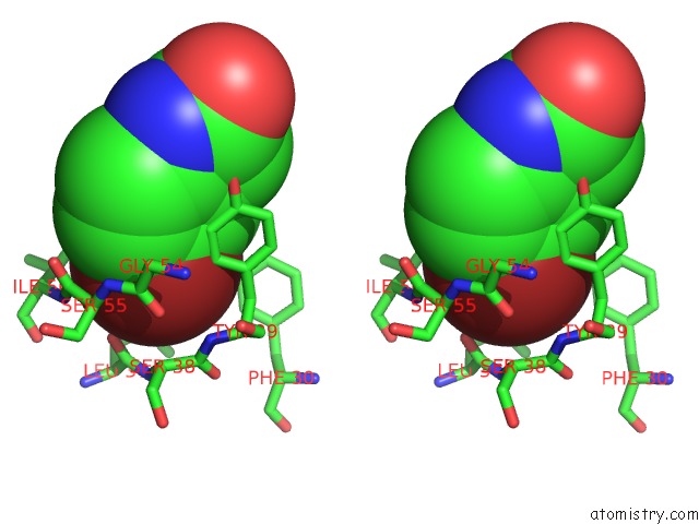
Bromine binding site 2 out of 4 in 6tvn
Go back to
Bromine binding site 2 out
of 4 in the Crystal Structure of 5-Bromoindoline-2,3-Dione Covalently Bound to the pH Domain of Bruton'S Tyrosine Kinase
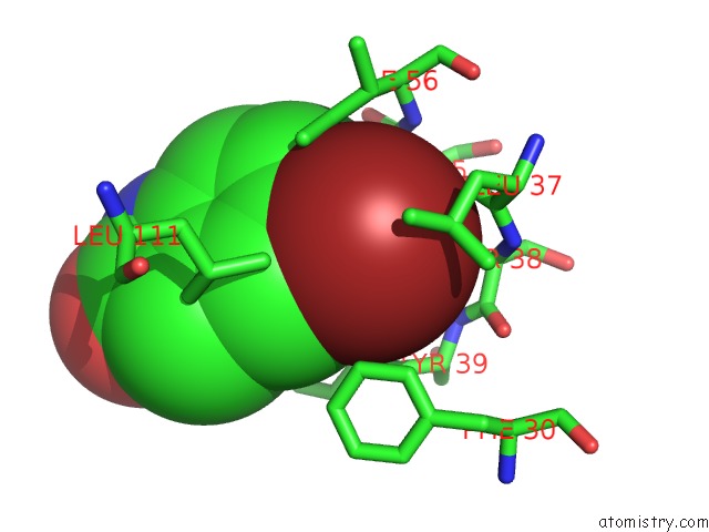
Mono view
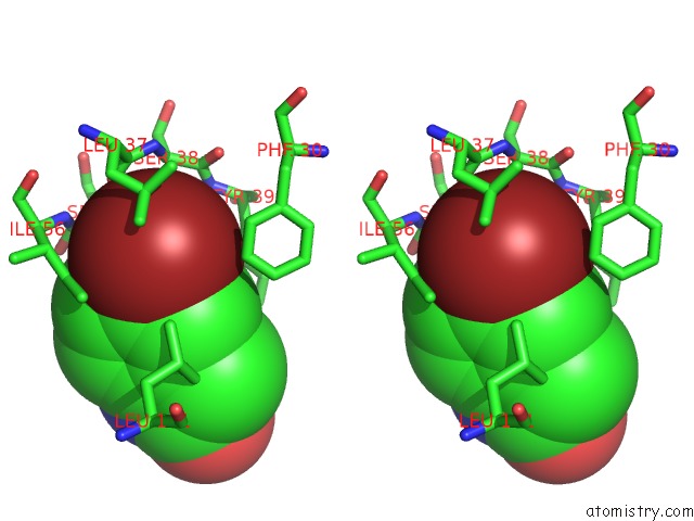
Stereo pair view
Mono view
Stereo pair view
A full contact list of Bromine with other atoms in the Br binding
site number 2 of Crystal Structure of 5-Bromoindoline-2,3-Dione Covalently Bound to the pH Domain of Bruton'S Tyrosine Kinase within 5.0Å range:
|
Bromine binding site 3 out of 4 in 6tvn
Go back to
Bromine binding site 3 out
of 4 in the Crystal Structure of 5-Bromoindoline-2,3-Dione Covalently Bound to the pH Domain of Bruton'S Tyrosine Kinase
Mono view

Stereo pair view
Mono view
Stereo pair view
A full contact list of Bromine with other atoms in the Br binding
site number 3 of Crystal Structure of 5-Bromoindoline-2,3-Dione Covalently Bound to the pH Domain of Bruton'S Tyrosine Kinase within 5.0Å range:
|
Bromine binding site 4 out of 4 in 6tvn
Go back to
Bromine binding site 4 out
of 4 in the Crystal Structure of 5-Bromoindoline-2,3-Dione Covalently Bound to the pH Domain of Bruton'S Tyrosine Kinase
Mono view
Stereo pair view
Mono view
Stereo pair view
A full contact list of Bromine with other atoms in the Br binding
site number 4 of Crystal Structure of 5-Bromoindoline-2,3-Dione Covalently Bound to the pH Domain of Bruton'S Tyrosine Kinase within 5.0Å range:
|
Reference:
P.Brear,
G.Fischer,
M.May,
T.Pantelejevs,
R.Mathieu,
M.Rossmann,
J.Wagstaff,
B.Blaszczyk,
M.Hyvonen.
Crystal Structure of 1-Methylindoline-2,3-Dione Covalently Bound to the pH Domain of Bruton'S Tyrosine Kinase Mutant R28C To Be Published.
Page generated: Thu Jul 11 02:54:32 2024
Last articles
As in 5IC5As in 5HWA
As in 5HRS
As in 5HRN
As in 5HRR
As in 5HRP
As in 5HR7
As in 5GM3
As in 5HLO
As in 5EZ3