Bromine »
PDB 7cud-7fl5 »
7fib »
Bromine in PDB 7fib: Crystal Structure of the Regulatory Domain of Acer in Acinetobacter Baumannii
Protein crystallography data
The structure of Crystal Structure of the Regulatory Domain of Acer in Acinetobacter Baumannii, PDB code: 7fib
was solved by
P.Chi,
L.Guo,
with X-Ray Crystallography technique. A brief refinement statistics is given in the table below:
| Resolution Low / High (Å) | 24.88 / 2.10 |
| Space group | P 65 |
| Cell size a, b, c (Å), α, β, γ (°) | 125.215, 125.215, 61.58, 90, 90, 120 |
| R / Rfree (%) | 20.5 / 23.1 |
Bromine Binding Sites:
The binding sites of Bromine atom in the Crystal Structure of the Regulatory Domain of Acer in Acinetobacter Baumannii
(pdb code 7fib). This binding sites where shown within
5.0 Angstroms radius around Bromine atom.
In total 5 binding sites of Bromine where determined in the Crystal Structure of the Regulatory Domain of Acer in Acinetobacter Baumannii, PDB code: 7fib:
Jump to Bromine binding site number: 1; 2; 3; 4; 5;
In total 5 binding sites of Bromine where determined in the Crystal Structure of the Regulatory Domain of Acer in Acinetobacter Baumannii, PDB code: 7fib:
Jump to Bromine binding site number: 1; 2; 3; 4; 5;
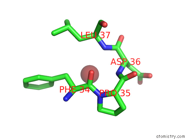
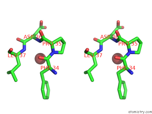
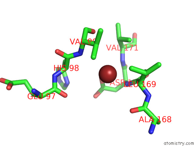
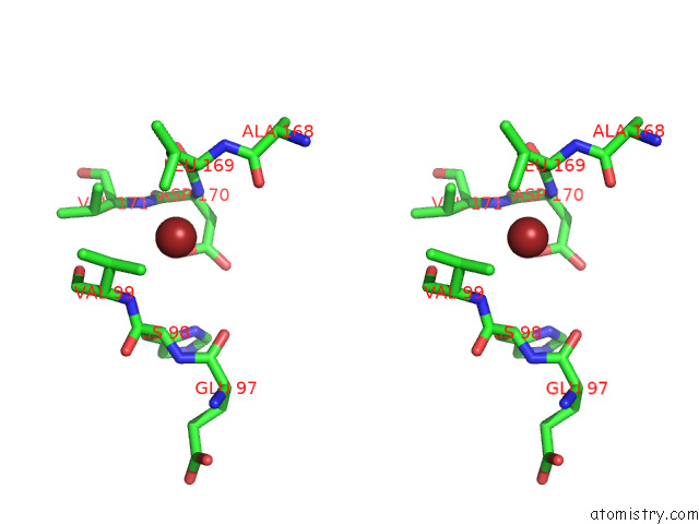
Bromine binding site 1 out of 5 in 7fib
Go back to
Bromine binding site 1 out
of 5 in the Crystal Structure of the Regulatory Domain of Acer in Acinetobacter Baumannii
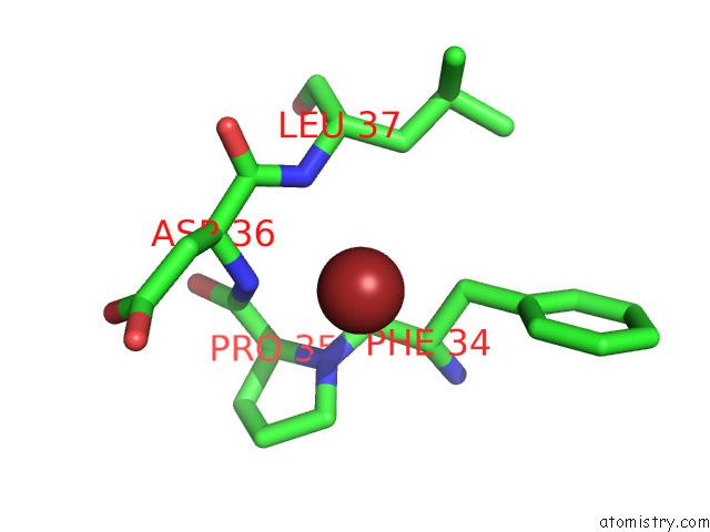
Mono view
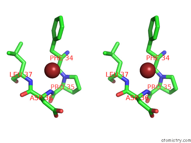
Stereo pair view
Mono view
Stereo pair view
A full contact list of Bromine with other atoms in the Br binding
site number 1 of Crystal Structure of the Regulatory Domain of Acer in Acinetobacter Baumannii within 5.0Å range:
|
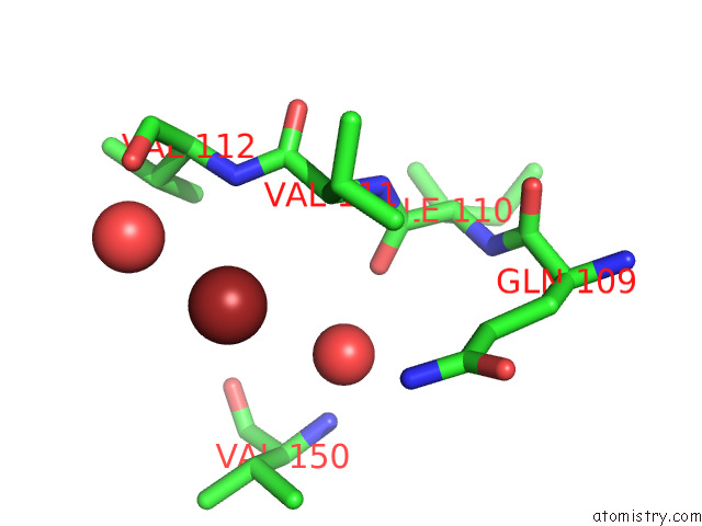
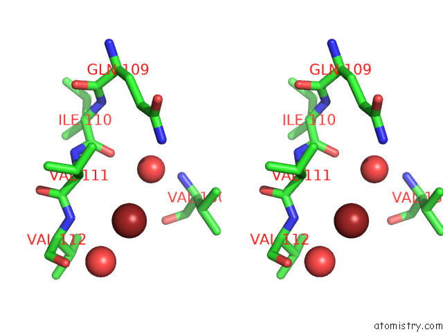
Bromine binding site 2 out of 5 in 7fib
Go back to
Bromine binding site 2 out
of 5 in the Crystal Structure of the Regulatory Domain of Acer in Acinetobacter Baumannii
Mono view
Stereo pair view
Mono view
Stereo pair view
A full contact list of Bromine with other atoms in the Br binding
site number 2 of Crystal Structure of the Regulatory Domain of Acer in Acinetobacter Baumannii within 5.0Å range:
|
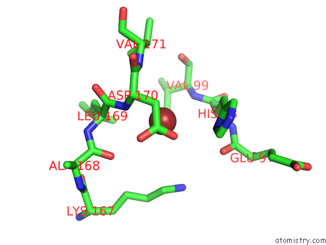
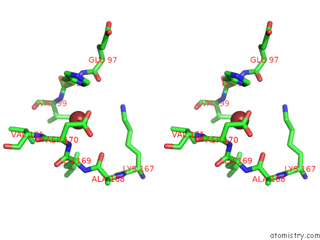
Bromine binding site 3 out of 5 in 7fib
Go back to
Bromine binding site 3 out
of 5 in the Crystal Structure of the Regulatory Domain of Acer in Acinetobacter Baumannii
Mono view
Stereo pair view
Mono view
Stereo pair view
A full contact list of Bromine with other atoms in the Br binding
site number 3 of Crystal Structure of the Regulatory Domain of Acer in Acinetobacter Baumannii within 5.0Å range:
|
Bromine binding site 4 out of 5 in 7fib
Go back to
Bromine binding site 4 out
of 5 in the Crystal Structure of the Regulatory Domain of Acer in Acinetobacter Baumannii
Mono view
Stereo pair view
Mono view
Stereo pair view
A full contact list of Bromine with other atoms in the Br binding
site number 4 of Crystal Structure of the Regulatory Domain of Acer in Acinetobacter Baumannii within 5.0Å range:
|
Bromine binding site 5 out of 5 in 7fib
Go back to
Bromine binding site 5 out
of 5 in the Crystal Structure of the Regulatory Domain of Acer in Acinetobacter Baumannii
Mono view
Stereo pair view
Mono view
Stereo pair view
A full contact list of Bromine with other atoms in the Br binding
site number 5 of Crystal Structure of the Regulatory Domain of Acer in Acinetobacter Baumannii within 5.0Å range:
|
Reference:
P.Chi,
L.Guo.
Crystal Structure of the Regulatory Domain of Acer in Acinetobacter Baumannii To Be Published.
Page generated: Thu Jul 11 03:48:57 2024
Last articles
Zn in 9J0NZn in 9J0O
Zn in 9J0P
Zn in 9FJX
Zn in 9EKB
Zn in 9C0F
Zn in 9CAH
Zn in 9CH0
Zn in 9CH3
Zn in 9CH1