Bromine »
PDB 7flh-7gd1 »
7flh »
Bromine in PDB 7flh: Pandda Analysis Group Deposition -- AAR2/Rnaseh in Complex with Fragment P05D03 From the F2X-Universal Library
Protein crystallography data
The structure of Pandda Analysis Group Deposition -- AAR2/Rnaseh in Complex with Fragment P05D03 From the F2X-Universal Library, PDB code: 7flh
was solved by
T.Barthel,
J.Wollenhaupt,
G.M.A.Lima,
M.C.Wahl,
M.S.Weiss,
with X-Ray Crystallography technique. A brief refinement statistics is given in the table below:
| Resolution Low / High (Å) | 44.28 / 1.67 |
| Space group | C 1 2 1 |
| Cell size a, b, c (Å), α, β, γ (°) | 86.679, 81.832, 93.348, 90, 108.26, 90 |
| R / Rfree (%) | 21 / 24.9 |
Bromine Binding Sites:
The binding sites of Bromine atom in the Pandda Analysis Group Deposition -- AAR2/Rnaseh in Complex with Fragment P05D03 From the F2X-Universal Library
(pdb code 7flh). This binding sites where shown within
5.0 Angstroms radius around Bromine atom.
In total only one binding site of Bromine was determined in the Pandda Analysis Group Deposition -- AAR2/Rnaseh in Complex with Fragment P05D03 From the F2X-Universal Library, PDB code: 7flh:
In total only one binding site of Bromine was determined in the Pandda Analysis Group Deposition -- AAR2/Rnaseh in Complex with Fragment P05D03 From the F2X-Universal Library, PDB code: 7flh:
Bromine binding site 1 out of 1 in 7flh
Go back to
Bromine binding site 1 out
of 1 in the Pandda Analysis Group Deposition -- AAR2/Rnaseh in Complex with Fragment P05D03 From the F2X-Universal Library
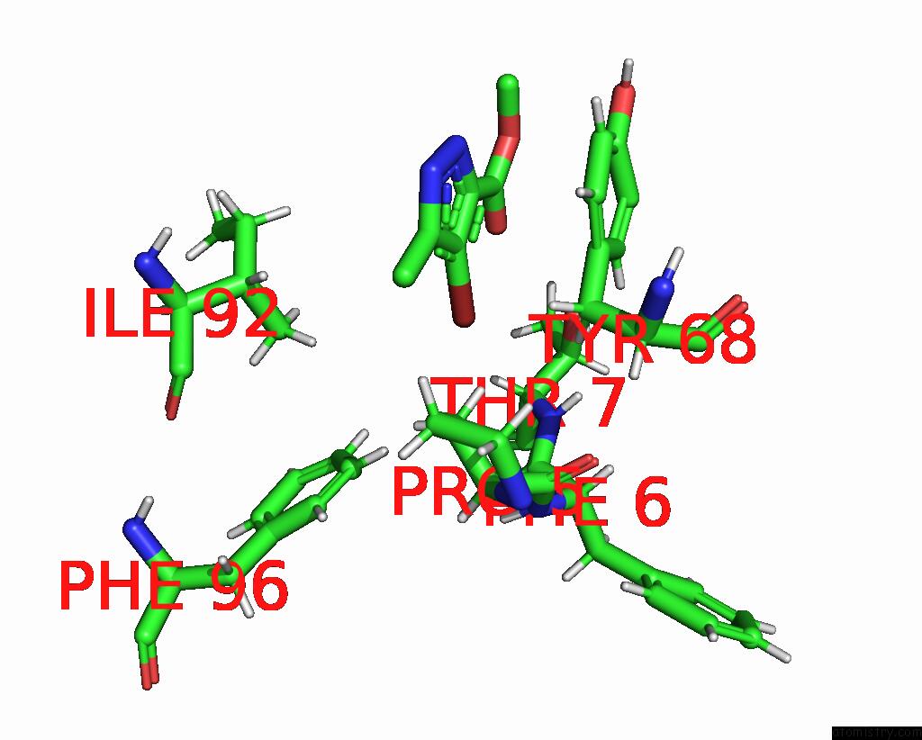
Mono view
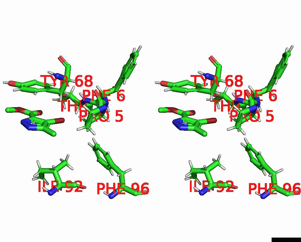
Stereo pair view
Mono view
Stereo pair view
A full contact list of Bromine with other atoms in the Br binding
site number 1 of Pandda Analysis Group Deposition -- AAR2/Rnaseh in Complex with Fragment P05D03 From the F2X-Universal Library within 5.0Å range:
|
Reference:
T.Barthel,
J.Wollenhaupt,
G.M.A.Lima,
M.C.Wahl,
M.S.Weiss.
Large-Scale Crystallographic Fragment Screening Expedites Compound Optimization and Identifies Putative Protein-Protein Interaction Sites. J.Med.Chem. V. 65 14630 2022.
ISSN: ISSN 0022-2623
PubMed: 36260741
DOI: 10.1021/ACS.JMEDCHEM.2C01165
Page generated: Mon Jul 7 11:15:46 2025
ISSN: ISSN 0022-2623
PubMed: 36260741
DOI: 10.1021/ACS.JMEDCHEM.2C01165
Last articles
Fe in 2YXOFe in 2YRS
Fe in 2YXC
Fe in 2YNM
Fe in 2YVJ
Fe in 2YP1
Fe in 2YU2
Fe in 2YU1
Fe in 2YQB
Fe in 2YOO