Bromine »
PDB 7gd2-7hhj »
7gyh »
Bromine in PDB 7gyh: Crystal Structure of HSP72 in Complex with Ligand 10 at 15.96 Mgy X- Ray Dose.
Protein crystallography data
The structure of Crystal Structure of HSP72 in Complex with Ligand 10 at 15.96 Mgy X- Ray Dose., PDB code: 7gyh
was solved by
M.Cabry,
M.J.Rodrigues,
Y.V.Le Bihan,
R.L.M.Van Montfort,
with X-Ray Crystallography technique. A brief refinement statistics is given in the table below:
| Resolution Low / High (Å) | 38.85 / 1.92 |
| Space group | P 21 21 21 |
| Cell size a, b, c (Å), α, β, γ (°) | 48.04, 89.89, 97.33, 90, 90, 90 |
| R / Rfree (%) | 17.7 / 23.7 |
Other elements in 7gyh:
The structure of Crystal Structure of HSP72 in Complex with Ligand 10 at 15.96 Mgy X- Ray Dose. also contains other interesting chemical elements:
| Magnesium | (Mg) | 1 atom |
Bromine Binding Sites:
The binding sites of Bromine atom in the Crystal Structure of HSP72 in Complex with Ligand 10 at 15.96 Mgy X- Ray Dose.
(pdb code 7gyh). This binding sites where shown within
5.0 Angstroms radius around Bromine atom.
In total only one binding site of Bromine was determined in the Crystal Structure of HSP72 in Complex with Ligand 10 at 15.96 Mgy X- Ray Dose., PDB code: 7gyh:
In total only one binding site of Bromine was determined in the Crystal Structure of HSP72 in Complex with Ligand 10 at 15.96 Mgy X- Ray Dose., PDB code: 7gyh:
Bromine binding site 1 out of 1 in 7gyh
Go back to
Bromine binding site 1 out
of 1 in the Crystal Structure of HSP72 in Complex with Ligand 10 at 15.96 Mgy X- Ray Dose.
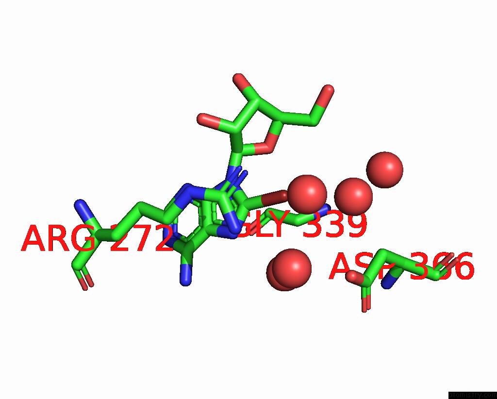
Mono view
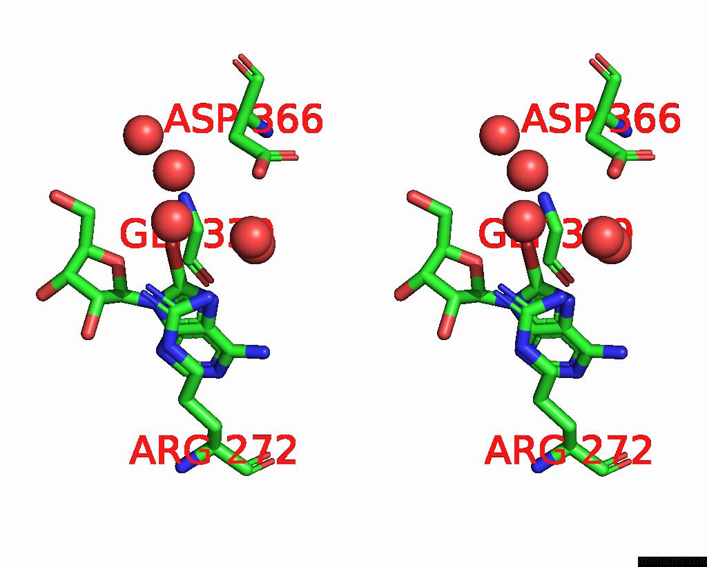
Stereo pair view
Mono view
Stereo pair view
A full contact list of Bromine with other atoms in the Br binding
site number 1 of Crystal Structure of HSP72 in Complex with Ligand 10 at 15.96 Mgy X- Ray Dose. within 5.0Å range:
|
Reference:
M.J.Rodrigues,
M.Cabry,
G.Collie,
M.Carter,
C.Mcandrew,
R.L.Owen,
B.R.Bellenie,
Y.-V.Le Bihan,
R.L.M.Van Montfort.
Specific Radiation Damage to Halogenated Inhibitors and Ligands in Protein-Ligand Crystal Structures J.Appl.Crystallogr. V. 57 1951 2024.
ISSN: ESSN 1600-5767
DOI: 10.1107/S1600576724010549
Page generated: Sun Dec 15 09:29:33 2024
ISSN: ESSN 1600-5767
DOI: 10.1107/S1600576724010549
Last articles
Zn in 9J0NZn in 9J0O
Zn in 9J0P
Zn in 9FJX
Zn in 9EKB
Zn in 9C0F
Zn in 9CAH
Zn in 9CH0
Zn in 9CH3
Zn in 9CH1