Bromine »
PDB 7qwb-7u5q »
7s3a »
Bromine in PDB 7s3a: Crystal Structure of Intact U2AF65 Rrm-Region Bound to Adml-C5 Oligonucleotide
Protein crystallography data
The structure of Crystal Structure of Intact U2AF65 Rrm-Region Bound to Adml-C5 Oligonucleotide, PDB code: 7s3a
was solved by
J.L.Jenkins,
S.Henderson,
C.L.Kielkopf,
with X-Ray Crystallography technique. A brief refinement statistics is given in the table below:
| Resolution Low / High (Å) | 37.76 / 1.48 |
| Space group | P 21 21 21 |
| Cell size a, b, c (Å), α, β, γ (°) | 43.245, 63.022, 77.491, 90, 90, 90 |
| R / Rfree (%) | 13.1 / 15.7 |
Other elements in 7s3a:
The structure of Crystal Structure of Intact U2AF65 Rrm-Region Bound to Adml-C5 Oligonucleotide also contains other interesting chemical elements:
| Sodium | (Na) | 3 atoms |
Bromine Binding Sites:
The binding sites of Bromine atom in the Crystal Structure of Intact U2AF65 Rrm-Region Bound to Adml-C5 Oligonucleotide
(pdb code 7s3a). This binding sites where shown within
5.0 Angstroms radius around Bromine atom.
In total only one binding site of Bromine was determined in the Crystal Structure of Intact U2AF65 Rrm-Region Bound to Adml-C5 Oligonucleotide, PDB code: 7s3a:
In total only one binding site of Bromine was determined in the Crystal Structure of Intact U2AF65 Rrm-Region Bound to Adml-C5 Oligonucleotide, PDB code: 7s3a:
Bromine binding site 1 out of 1 in 7s3a
Go back to
Bromine binding site 1 out
of 1 in the Crystal Structure of Intact U2AF65 Rrm-Region Bound to Adml-C5 Oligonucleotide
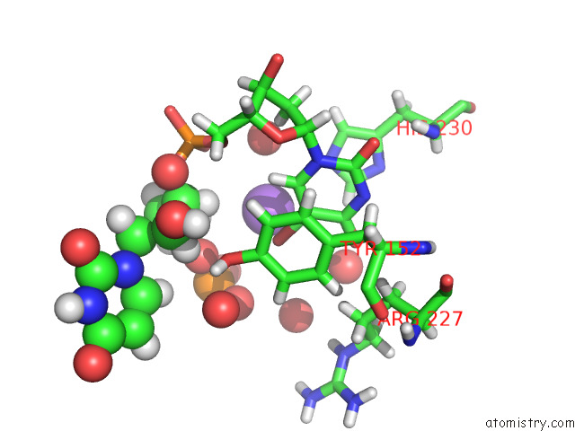
Mono view
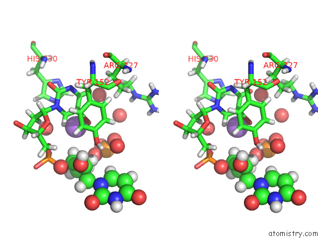
Stereo pair view
Mono view
Stereo pair view
A full contact list of Bromine with other atoms in the Br binding
site number 1 of Crystal Structure of Intact U2AF65 Rrm-Region Bound to Adml-C5 Oligonucleotide within 5.0Å range:
|
Reference:
E.Glasser,
D.Maji,
G.Biancon,
A.M.K.Puthenpeedikakkal,
C.E.Cavender,
T.Tebaldi,
J.L.Jenkins,
D.H.Mathews,
S.Halene,
C.L.Kielkopf.
Pre-Mrna Splicing Factor U2AF2 Recognizes Distinct Conformations of Nucleotide Variants at the Center of the Pre-Mrna Splice Site Signal. Nucleic Acids Res. V. 50 5299 2022.
ISSN: ESSN 1362-4962
PubMed: 35524551
DOI: 10.1093/NAR/GKAC287
Page generated: Mon Jul 7 11:47:39 2025
ISSN: ESSN 1362-4962
PubMed: 35524551
DOI: 10.1093/NAR/GKAC287
Last articles
Cl in 8CHMCl in 8CHY
Cl in 8CHR
Cl in 8CHQ
Cl in 8CHP
Cl in 8CHL
Cl in 8CHJ
Cl in 8CHN
Cl in 8CHK
Cl in 8CHI