Bromine »
PDB 8gew-8k2l »
8joi »
Bromine in PDB 8joi: Crystal Structure of Bel-1 E166A Mutant.
Protein crystallography data
The structure of Crystal Structure of Bel-1 E166A Mutant., PDB code: 8joi
was solved by
K.Dhankhar,
S.Hazra,
with X-Ray Crystallography technique. A brief refinement statistics is given in the table below:
| Resolution Low / High (Å) | 26.61 / 1.70 |
| Space group | P 6 |
| Cell size a, b, c (Å), α, β, γ (°) | 123.498, 123.498, 70.644, 90, 90, 120 |
| R / Rfree (%) | 16.1 / 22.5 |
Bromine Binding Sites:
The binding sites of Bromine atom in the Crystal Structure of Bel-1 E166A Mutant.
(pdb code 8joi). This binding sites where shown within
5.0 Angstroms radius around Bromine atom.
In total only one binding site of Bromine was determined in the Crystal Structure of Bel-1 E166A Mutant., PDB code: 8joi:
In total only one binding site of Bromine was determined in the Crystal Structure of Bel-1 E166A Mutant., PDB code: 8joi:
Bromine binding site 1 out of 1 in 8joi
Go back to
Bromine binding site 1 out
of 1 in the Crystal Structure of Bel-1 E166A Mutant.
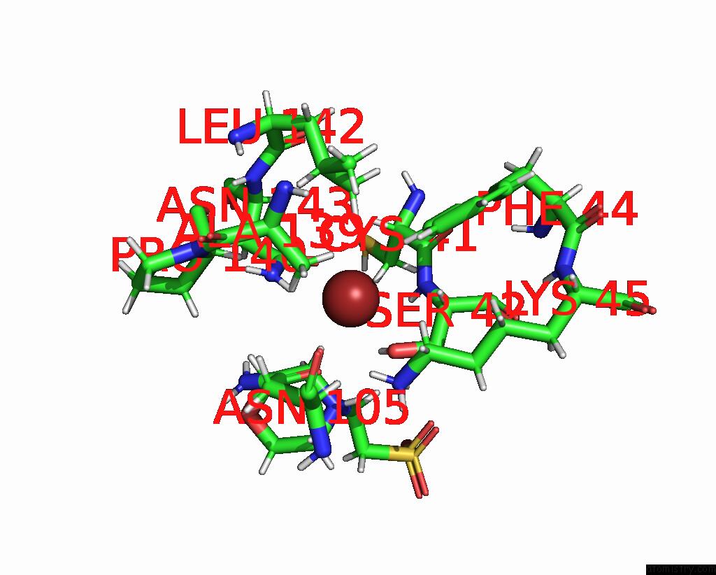
Mono view
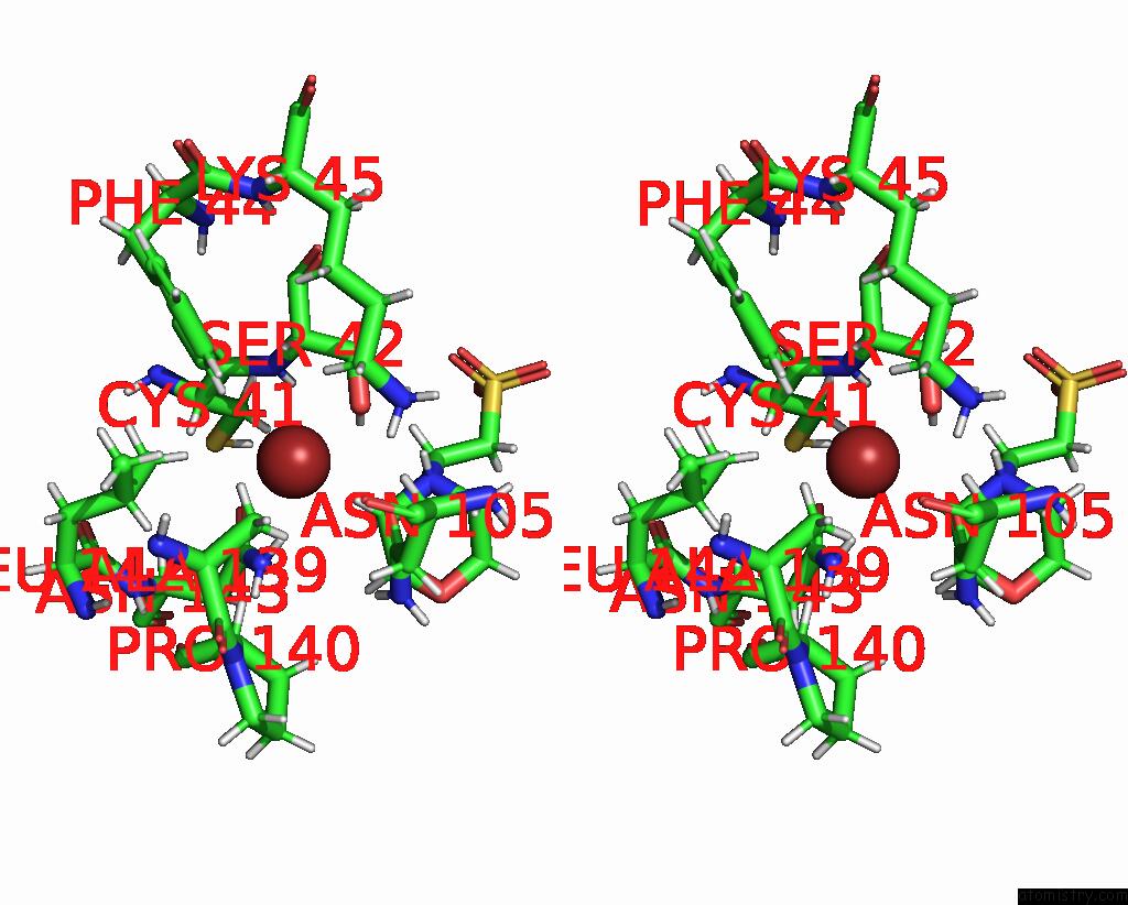
Stereo pair view
Mono view
Stereo pair view
A full contact list of Bromine with other atoms in the Br binding
site number 1 of Crystal Structure of Bel-1 E166A Mutant. within 5.0Å range:
|
Reference:
K.Dhankhar,
S.Hazra.
Crystal Structure of Bel-1 E166A Mutant To Be Published.
Page generated: Mon Jul 7 12:25:57 2025
Last articles
Cl in 5OGHCl in 5OHE
Cl in 5OHI
Cl in 5OHH
Cl in 5OGC
Cl in 5OG9
Cl in 5OFW
Cl in 5OFU
Cl in 5OFS
Cl in 5OG7