Bromine »
PDB 8gih-8k2m »
8k2l »
Bromine in PDB 8k2l: Crystal Structure of Group 4 Monosaccharide-Releasing Beta-N- Acetylgalactosaminidase NGAP2 From Paenibacillus Sp. TS12, Apo Form
Protein crystallography data
The structure of Crystal Structure of Group 4 Monosaccharide-Releasing Beta-N- Acetylgalactosaminidase NGAP2 From Paenibacillus Sp. TS12, Apo Form, PDB code: 8k2l
was solved by
T.Sumida,
S.Fushinobu,
with X-Ray Crystallography technique. A brief refinement statistics is given in the table below:
| Resolution Low / High (Å) | 46.70 / 1.95 |
| Space group | C 1 2 1 |
| Cell size a, b, c (Å), α, β, γ (°) | 119.116, 60.375, 100.285, 90, 120.08, 90 |
| R / Rfree (%) | 14.8 / 19.5 |
Bromine Binding Sites:
The binding sites of Bromine atom in the Crystal Structure of Group 4 Monosaccharide-Releasing Beta-N- Acetylgalactosaminidase NGAP2 From Paenibacillus Sp. TS12, Apo Form
(pdb code 8k2l). This binding sites where shown within
5.0 Angstroms radius around Bromine atom.
In total only one binding site of Bromine was determined in the Crystal Structure of Group 4 Monosaccharide-Releasing Beta-N- Acetylgalactosaminidase NGAP2 From Paenibacillus Sp. TS12, Apo Form, PDB code: 8k2l:
In total only one binding site of Bromine was determined in the Crystal Structure of Group 4 Monosaccharide-Releasing Beta-N- Acetylgalactosaminidase NGAP2 From Paenibacillus Sp. TS12, Apo Form, PDB code: 8k2l:
Bromine binding site 1 out of 1 in 8k2l
Go back to
Bromine binding site 1 out
of 1 in the Crystal Structure of Group 4 Monosaccharide-Releasing Beta-N- Acetylgalactosaminidase NGAP2 From Paenibacillus Sp. TS12, Apo Form
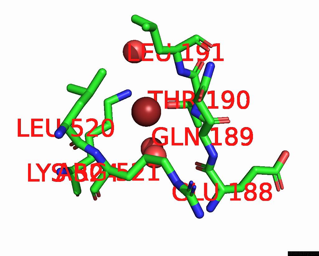
Mono view
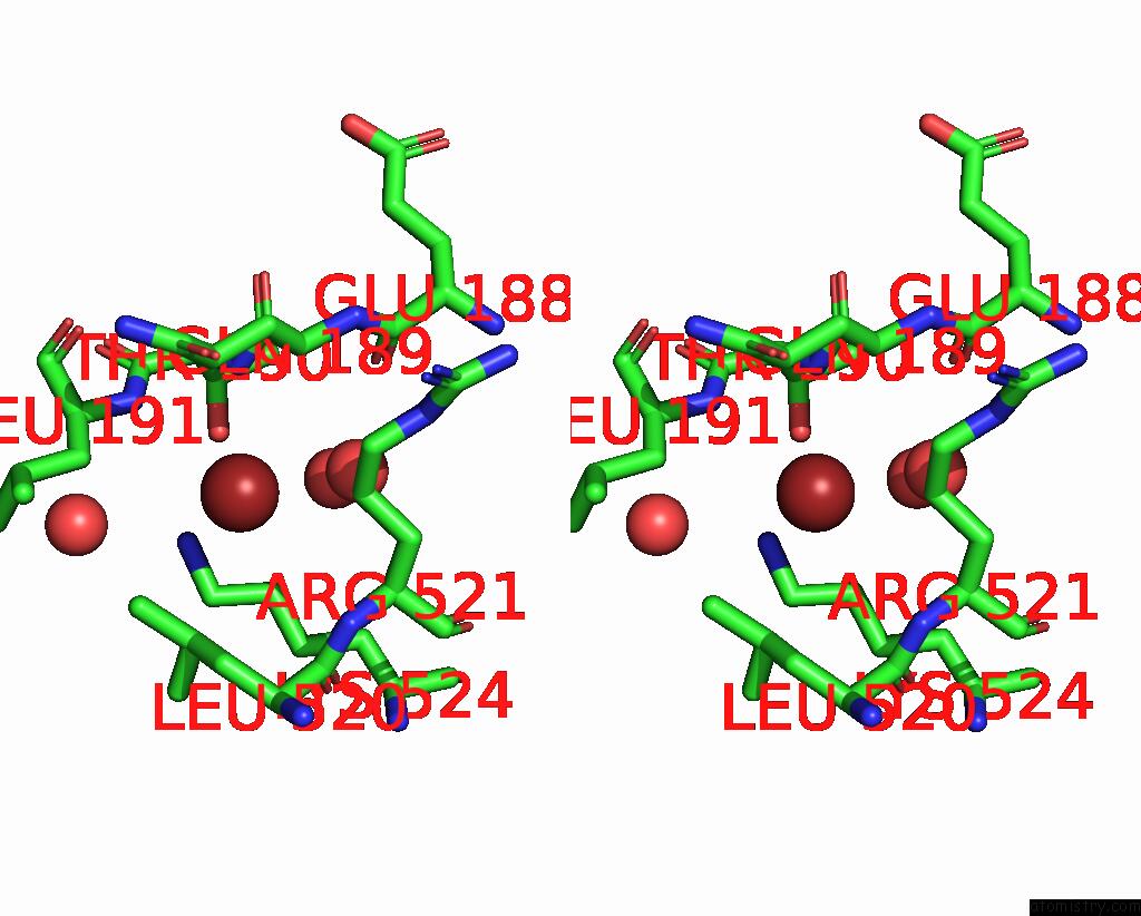
Stereo pair view
Mono view
Stereo pair view
A full contact list of Bromine with other atoms in the Br binding
site number 1 of Crystal Structure of Group 4 Monosaccharide-Releasing Beta-N- Acetylgalactosaminidase NGAP2 From Paenibacillus Sp. TS12, Apo Form within 5.0Å range:
|
Reference:
T.Sumida,
S.Hiraoka,
K.Usui,
A.Ishiwata,
T.Sengoku,
K.A.Stubbs,
K.Tanaka,
S.Deguchi,
S.Fushinobu,
T.Nunoura.
Genetic and Functional Diversity of Beta-N-Acetylgalactosamine Residue-Targeting Glycosidases Expanded By Deep-Sea Metagenome To Be Published.
Page generated: Thu Jul 11 05:23:33 2024
Last articles
Zn in 9MJ5Zn in 9HNW
Zn in 9G0L
Zn in 9FNE
Zn in 9DZN
Zn in 9E0I
Zn in 9D32
Zn in 9DAK
Zn in 8ZXC
Zn in 8ZUF