Bromine »
PDB 1e7b-1ih6 »
1hd2 »
Bromine in PDB 1hd2: Human Peroxiredoxin 5
Protein crystallography data
The structure of Human Peroxiredoxin 5, PDB code: 1hd2
was solved by
J.P.Declercq,
C.Evrard,
with X-Ray Crystallography technique. A brief refinement statistics is given in the table below:
| Resolution Low / High (Å) | 16.00 / 1.50 |
| Space group | P 41 21 2 |
| Cell size a, b, c (Å), α, β, γ (°) | 66.607, 66.607, 123.327, 90.00, 90.00, 90.00 |
| R / Rfree (%) | 13.3 / 16.5 |
Bromine Binding Sites:
The binding sites of Bromine atom in the Human Peroxiredoxin 5
(pdb code 1hd2). This binding sites where shown within
5.0 Angstroms radius around Bromine atom.
In total 5 binding sites of Bromine where determined in the Human Peroxiredoxin 5, PDB code: 1hd2:
Jump to Bromine binding site number: 1; 2; 3; 4; 5;
In total 5 binding sites of Bromine where determined in the Human Peroxiredoxin 5, PDB code: 1hd2:
Jump to Bromine binding site number: 1; 2; 3; 4; 5;
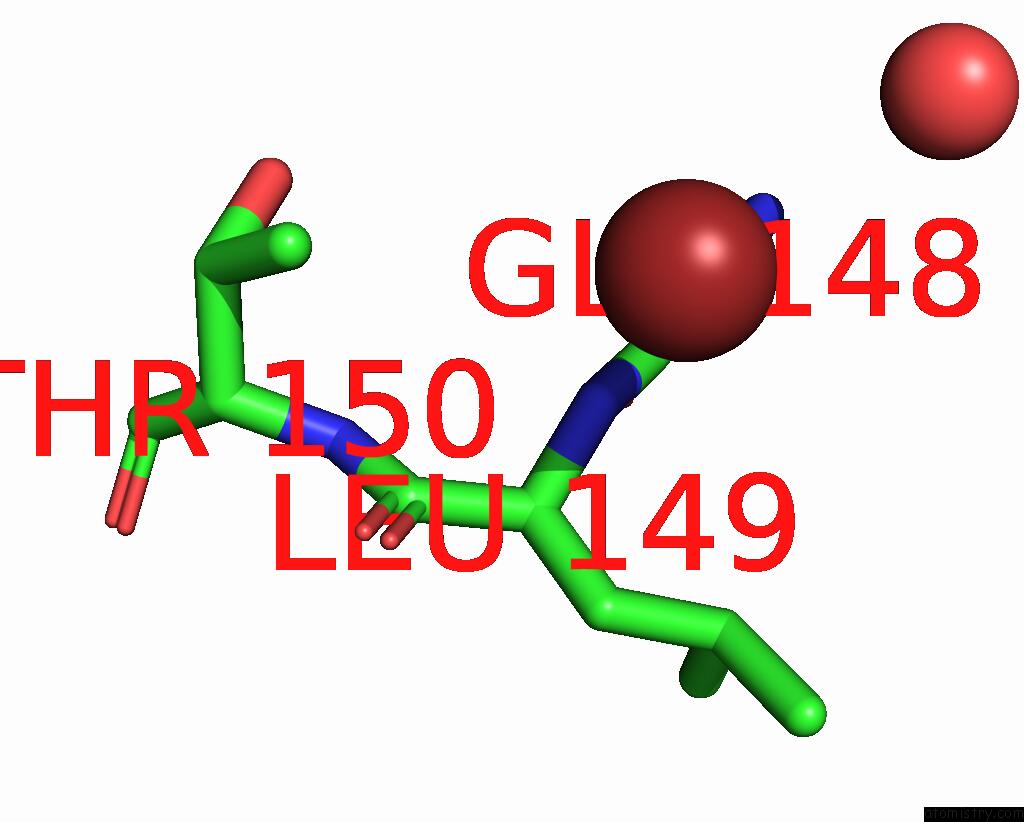
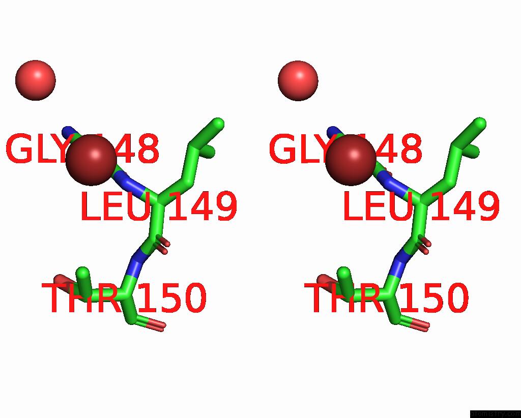
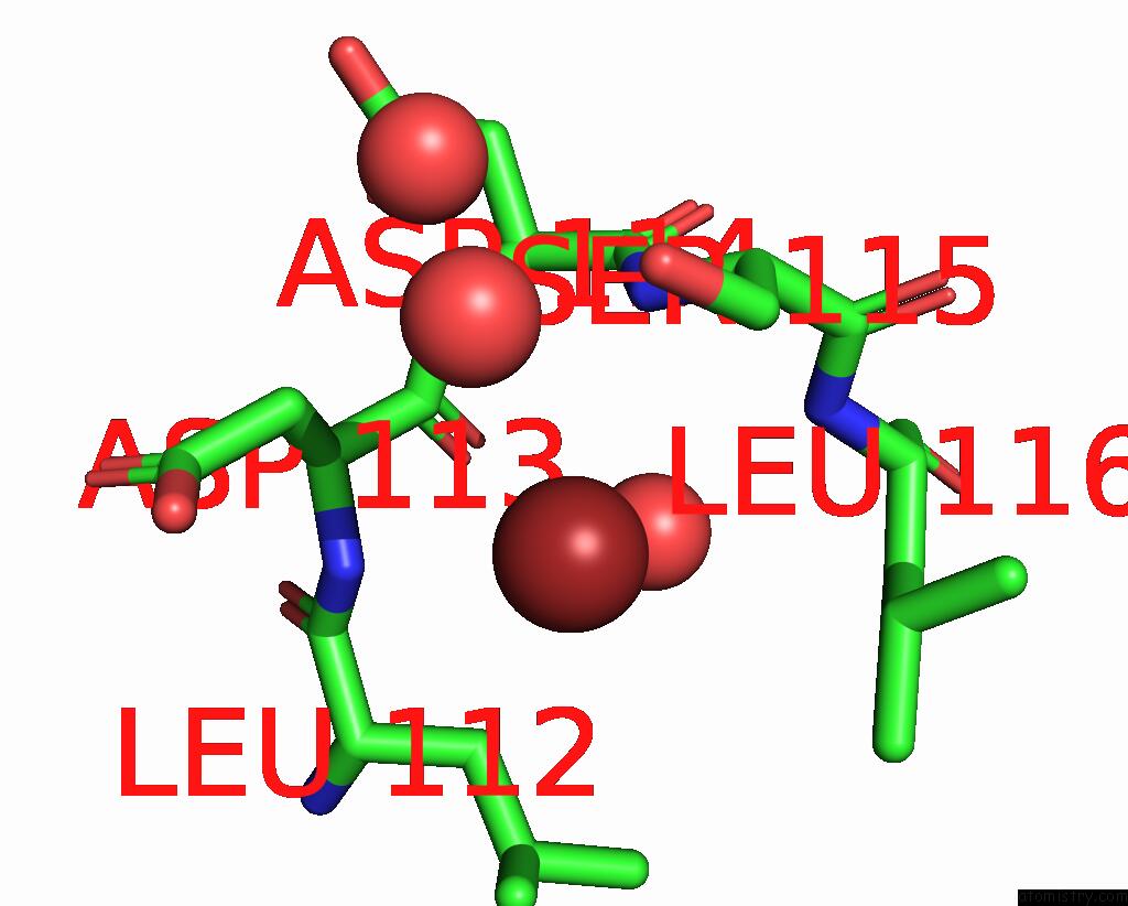
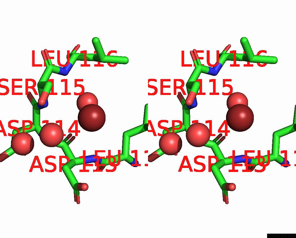
Bromine binding site 1 out of 5 in 1hd2
Go back to
Bromine binding site 1 out
of 5 in the Human Peroxiredoxin 5
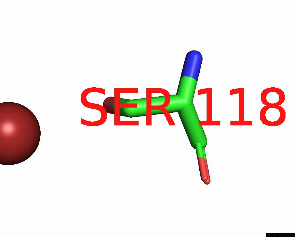
Mono view
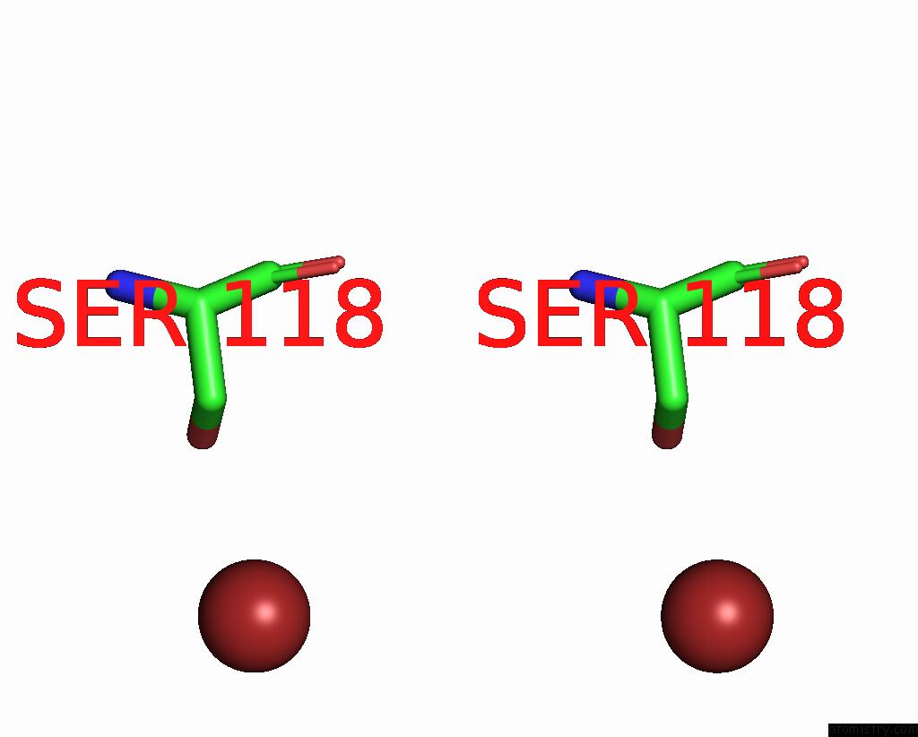
Stereo pair view
Mono view
Stereo pair view
A full contact list of Bromine with other atoms in the Br binding
site number 1 of Human Peroxiredoxin 5 within 5.0Å range:
|
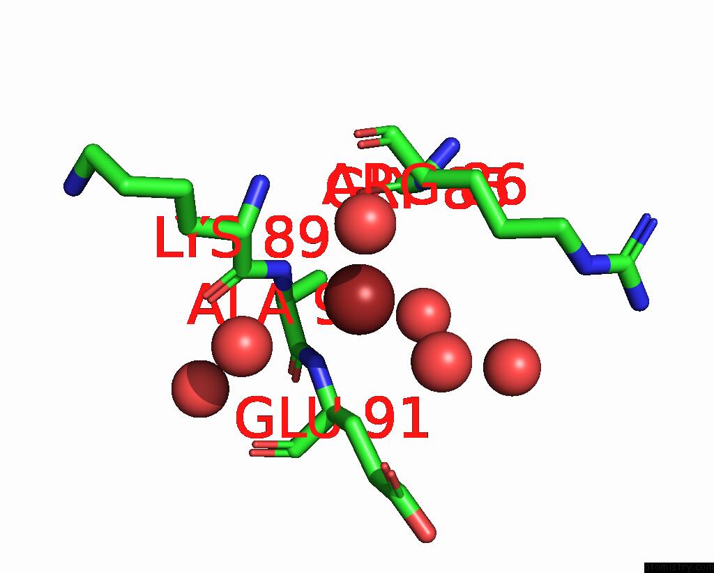
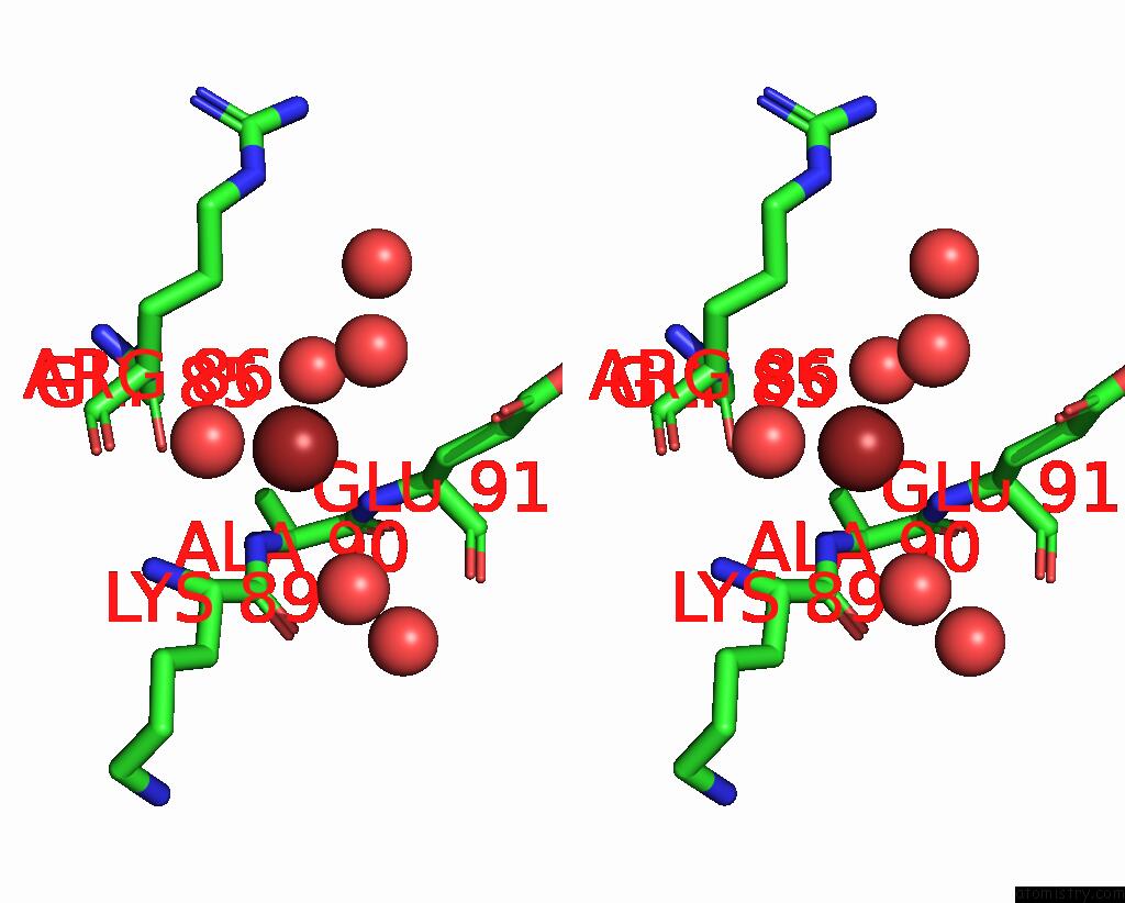
Bromine binding site 2 out of 5 in 1hd2
Go back to
Bromine binding site 2 out
of 5 in the Human Peroxiredoxin 5
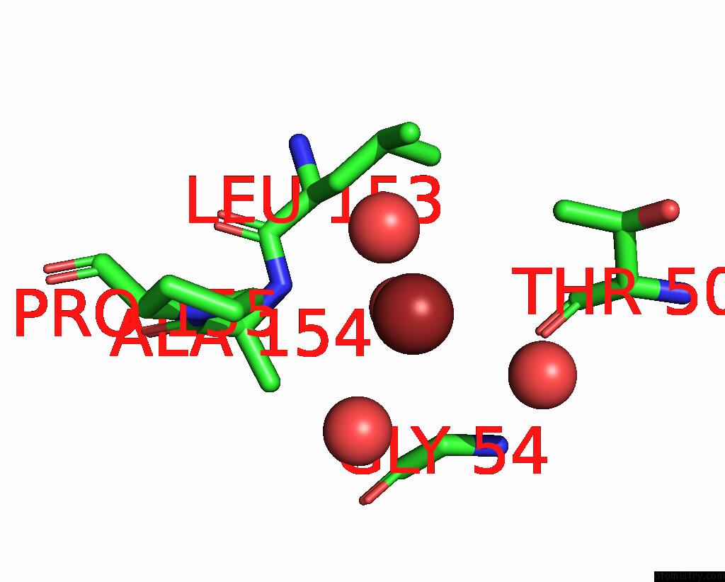
Mono view
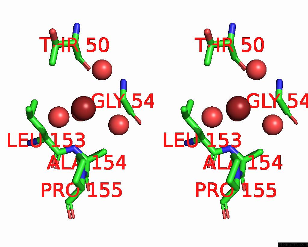
Stereo pair view
Mono view
Stereo pair view
A full contact list of Bromine with other atoms in the Br binding
site number 2 of Human Peroxiredoxin 5 within 5.0Å range:
|
Bromine binding site 3 out of 5 in 1hd2
Go back to
Bromine binding site 3 out
of 5 in the Human Peroxiredoxin 5
Mono view
Stereo pair view
Mono view
Stereo pair view
A full contact list of Bromine with other atoms in the Br binding
site number 3 of Human Peroxiredoxin 5 within 5.0Å range:
|
Bromine binding site 4 out of 5 in 1hd2
Go back to
Bromine binding site 4 out
of 5 in the Human Peroxiredoxin 5
Mono view
Stereo pair view
Mono view
Stereo pair view
A full contact list of Bromine with other atoms in the Br binding
site number 4 of Human Peroxiredoxin 5 within 5.0Å range:
|
Bromine binding site 5 out of 5 in 1hd2
Go back to
Bromine binding site 5 out
of 5 in the Human Peroxiredoxin 5
Mono view
Stereo pair view
Mono view
Stereo pair view
A full contact list of Bromine with other atoms in the Br binding
site number 5 of Human Peroxiredoxin 5 within 5.0Å range:
|
Reference:
J.P.Declercq,
C.Evrard,
A.Clippe,
D.V.Stricht,
A.Bernard,
B.Knoops.
Crystal Structure of Human Peroxiredoxin 5, A Novel Type of Mammalian Peroxiredoxin at 1.5 A Resolution. J.Mol.Biol. V. 311 751 2001.
ISSN: ISSN 0022-2836
PubMed: 11518528
DOI: 10.1006/JMBI.2001.4853
Page generated: Mon Jul 7 03:17:08 2025
ISSN: ISSN 0022-2836
PubMed: 11518528
DOI: 10.1006/JMBI.2001.4853
Last articles
Na in 7DNBNa in 7DLN
Na in 7DFO
Na in 7DLH
Na in 7DJQ
Na in 7DJK
Na in 7DKM
Na in 7DJM
Na in 7DJL
Na in 7DJC