Bromine »
PDB 1iha-1m9r »
1kfo »
Bromine in PDB 1kfo: Crystal Structure of An Rna Helix Recognized By A Zinc- Finger Protein: An 18 Base Pair Duplex at 1.6 Resolution
Protein crystallography data
The structure of Crystal Structure of An Rna Helix Recognized By A Zinc- Finger Protein: An 18 Base Pair Duplex at 1.6 Resolution, PDB code: 1kfo
was solved by
S.Lima,
J.Hildenbrand,
A.Korostelev,
S.Hattman,
H.Li,
with X-Ray Crystallography technique. A brief refinement statistics is given in the table below:
| Resolution Low / High (Å) | 16.69 / 1.60 |
| Space group | P 63 2 2 |
| Cell size a, b, c (Å), α, β, γ (°) | 45.336, 45.336, 95.129, 90.00, 90.00, 120.00 |
| R / Rfree (%) | 26 / 28.8 |
Bromine Binding Sites:
The binding sites of Bromine atom in the Crystal Structure of An Rna Helix Recognized By A Zinc- Finger Protein: An 18 Base Pair Duplex at 1.6 Resolution
(pdb code 1kfo). This binding sites where shown within
5.0 Angstroms radius around Bromine atom.
In total only one binding site of Bromine was determined in the Crystal Structure of An Rna Helix Recognized By A Zinc- Finger Protein: An 18 Base Pair Duplex at 1.6 Resolution, PDB code: 1kfo:
In total only one binding site of Bromine was determined in the Crystal Structure of An Rna Helix Recognized By A Zinc- Finger Protein: An 18 Base Pair Duplex at 1.6 Resolution, PDB code: 1kfo:
Bromine binding site 1 out of 1 in 1kfo
Go back to
Bromine binding site 1 out
of 1 in the Crystal Structure of An Rna Helix Recognized By A Zinc- Finger Protein: An 18 Base Pair Duplex at 1.6 Resolution
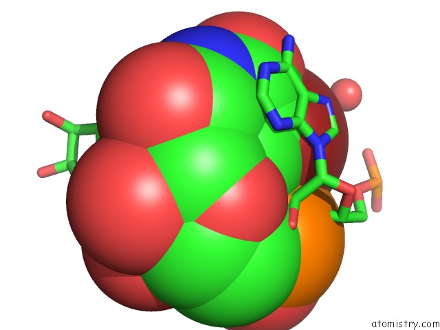
Mono view
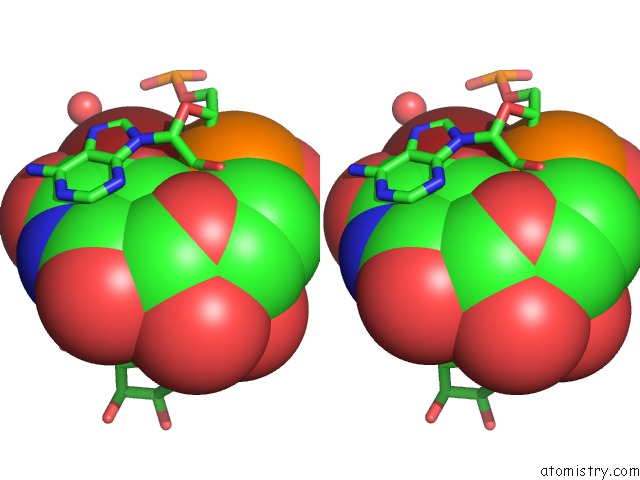
Stereo pair view
Mono view
Stereo pair view
A full contact list of Bromine with other atoms in the Br binding
site number 1 of Crystal Structure of An Rna Helix Recognized By A Zinc- Finger Protein: An 18 Base Pair Duplex at 1.6 Resolution within 5.0Å range:
|
Reference:
S.Lima,
J.Hildenbrand,
A.Korostelev,
S.Hattman,
H.Li.
Crystal Structure of An Rna Helix Recognized By A Zinc-Finger Protein: An 18-Bp Duplex at 1.6 A Resolution. Rna V. 8 924 2002.
ISSN: ISSN 1355-8382
PubMed: 12166647
DOI: 10.1017/S1355838202028893
Page generated: Mon Jul 7 03:22:37 2025
ISSN: ISSN 1355-8382
PubMed: 12166647
DOI: 10.1017/S1355838202028893
Last articles
Mn in 9LJUMn in 9LJW
Mn in 9LJS
Mn in 9LJR
Mn in 9LJT
Mn in 9LJV
Mg in 9UA2
Mg in 9R96
Mg in 9VM1
Mg in 9P01