Bromine »
PDB 3gw6-3jtk »
3ivn »
Bromine in PDB 3ivn: Structure of the U65C Mutant A-Riboswitch Aptamer From the Bacillus Subtilis Pbue Operon
Protein crystallography data
The structure of Structure of the U65C Mutant A-Riboswitch Aptamer From the Bacillus Subtilis Pbue Operon, PDB code: 3ivn
was solved by
V.Delfosse,
P.Dagenais,
D.Chausse,
G.Di Tomasso,
P.Legault,
with X-Ray Crystallography technique. A brief refinement statistics is given in the table below:
| Resolution Low / High (Å) | 26.04 / 2.80 |
| Space group | C 1 2 1 |
| Cell size a, b, c (Å), α, β, γ (°) | 124.930, 46.330, 87.240, 90.00, 120.04, 90.00 |
| R / Rfree (%) | 22.7 / 27.7 |
Other elements in 3ivn:
The structure of Structure of the U65C Mutant A-Riboswitch Aptamer From the Bacillus Subtilis Pbue Operon also contains other interesting chemical elements:
| Magnesium | (Mg) | 9 atoms |
Bromine Binding Sites:
The binding sites of Bromine atom in the Structure of the U65C Mutant A-Riboswitch Aptamer From the Bacillus Subtilis Pbue Operon
(pdb code 3ivn). This binding sites where shown within
5.0 Angstroms radius around Bromine atom.
In total 2 binding sites of Bromine where determined in the Structure of the U65C Mutant A-Riboswitch Aptamer From the Bacillus Subtilis Pbue Operon, PDB code: 3ivn:
Jump to Bromine binding site number: 1; 2;
In total 2 binding sites of Bromine where determined in the Structure of the U65C Mutant A-Riboswitch Aptamer From the Bacillus Subtilis Pbue Operon, PDB code: 3ivn:
Jump to Bromine binding site number: 1; 2;
Bromine binding site 1 out of 2 in 3ivn
Go back to
Bromine binding site 1 out
of 2 in the Structure of the U65C Mutant A-Riboswitch Aptamer From the Bacillus Subtilis Pbue Operon
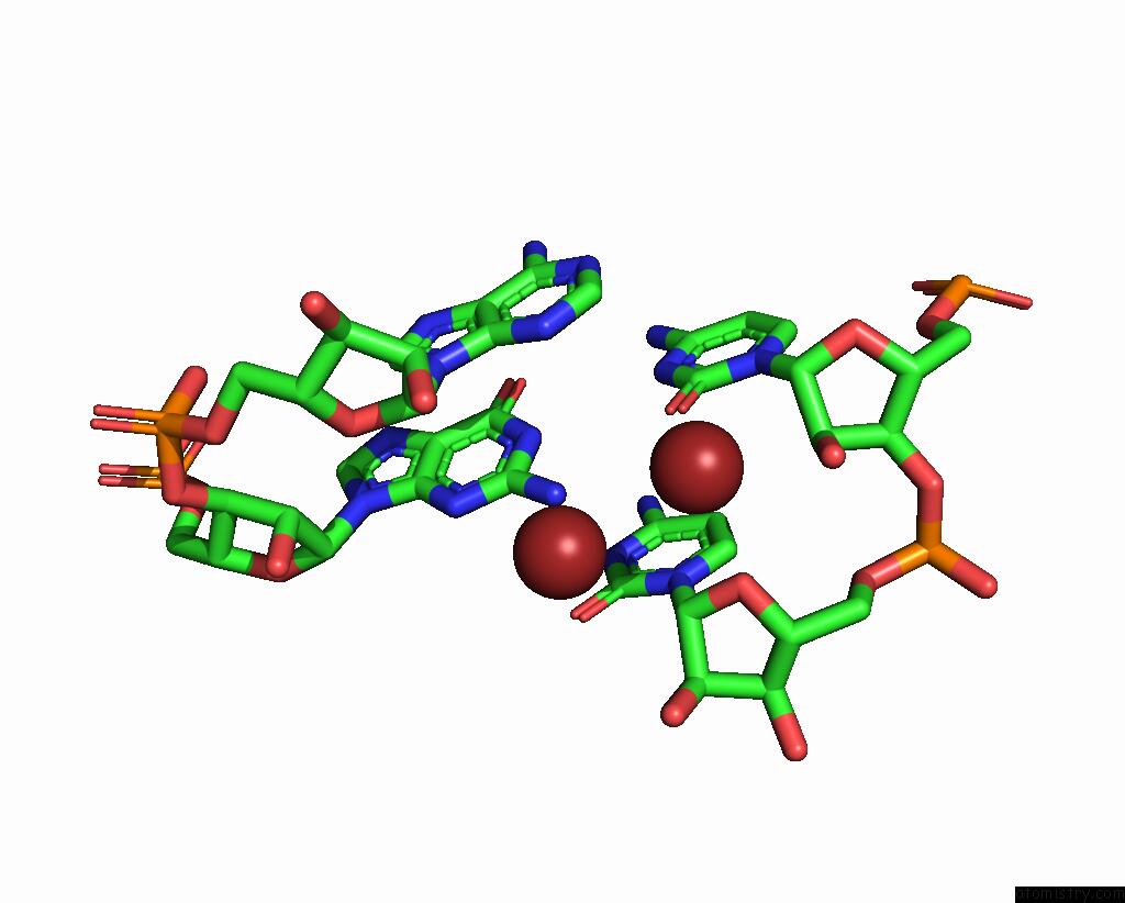
Mono view
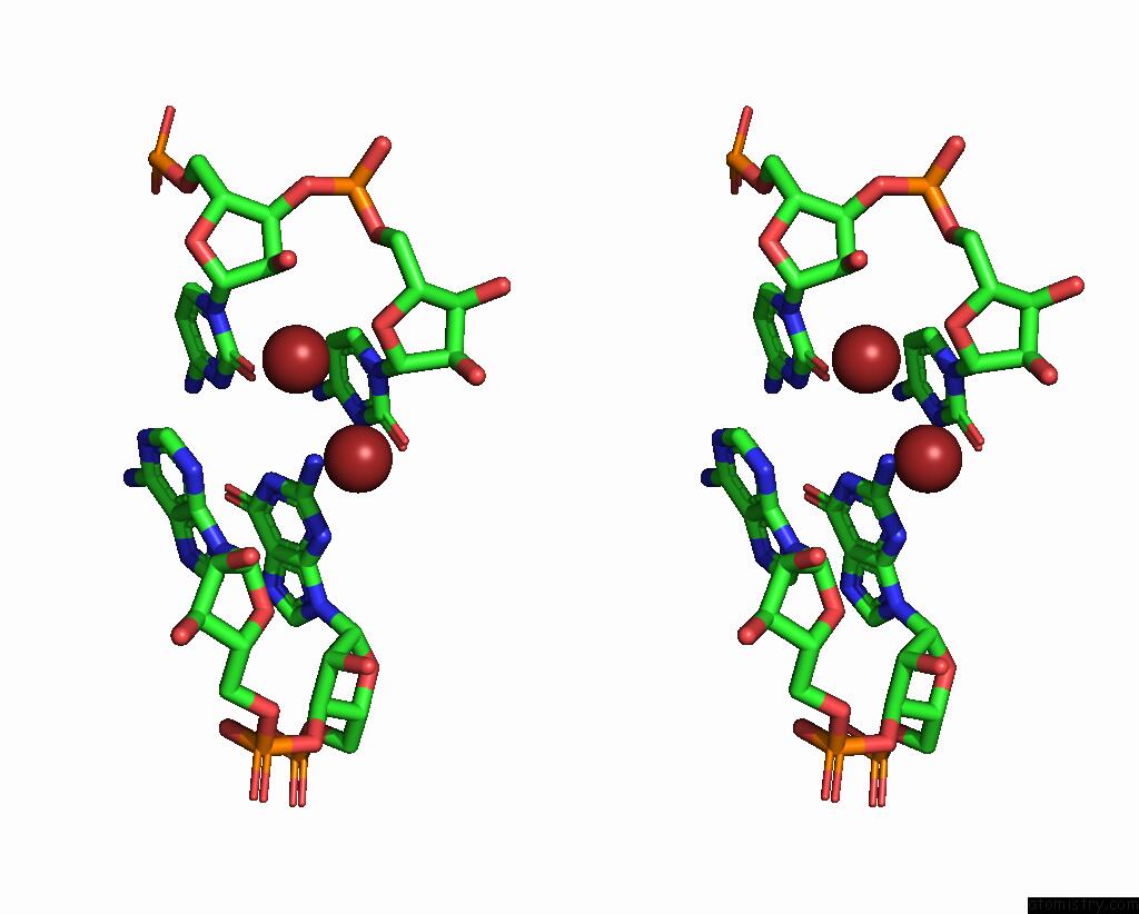
Stereo pair view
Mono view
Stereo pair view
A full contact list of Bromine with other atoms in the Br binding
site number 1 of Structure of the U65C Mutant A-Riboswitch Aptamer From the Bacillus Subtilis Pbue Operon within 5.0Å range:
|
Bromine binding site 2 out of 2 in 3ivn
Go back to
Bromine binding site 2 out
of 2 in the Structure of the U65C Mutant A-Riboswitch Aptamer From the Bacillus Subtilis Pbue Operon
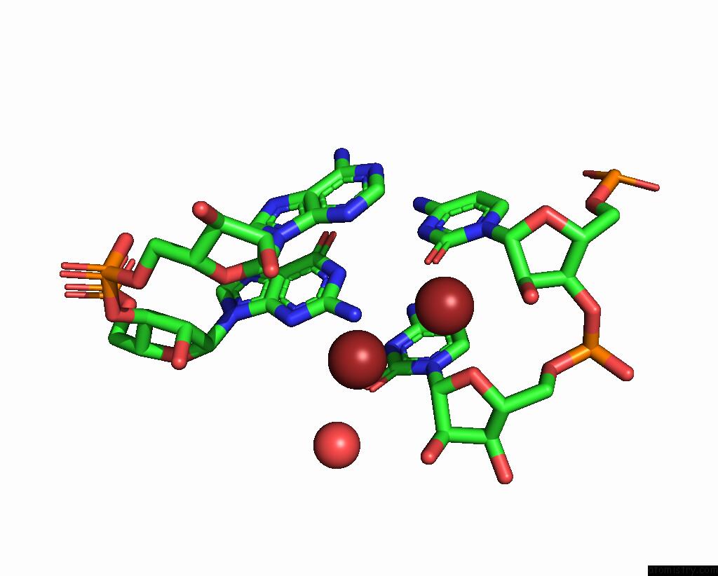
Mono view
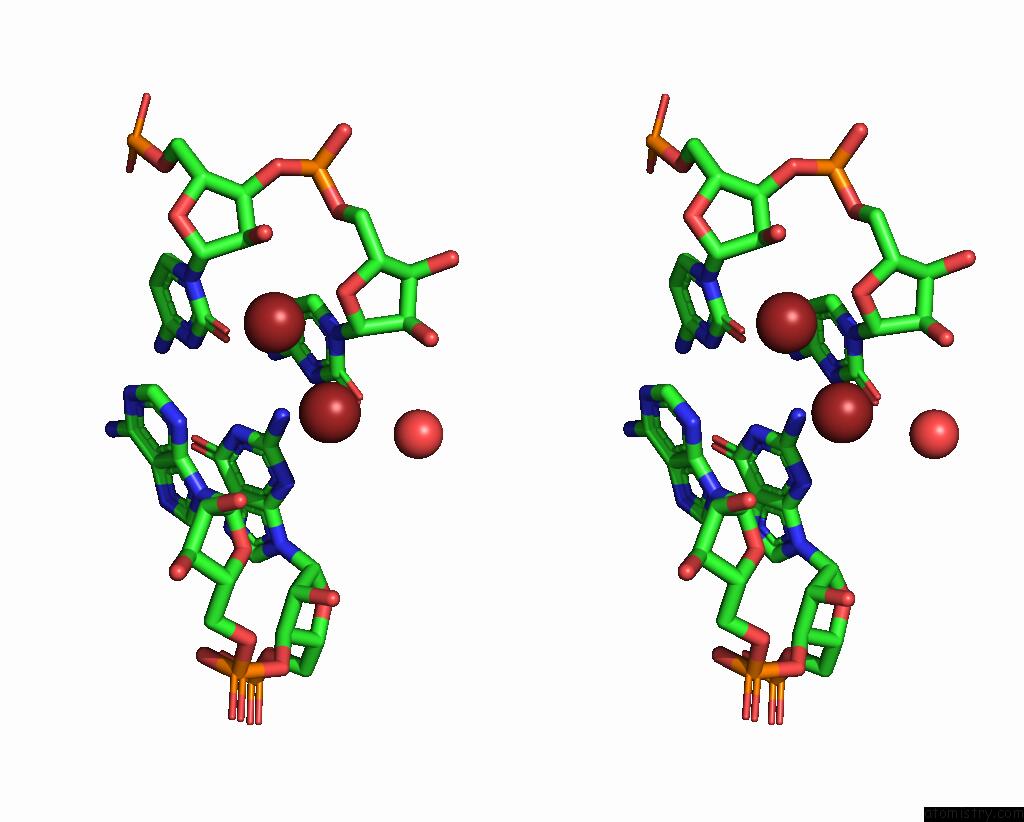
Stereo pair view
Mono view
Stereo pair view
A full contact list of Bromine with other atoms in the Br binding
site number 2 of Structure of the U65C Mutant A-Riboswitch Aptamer From the Bacillus Subtilis Pbue Operon within 5.0Å range:
|
Reference:
V.Delfosse,
P.Bouchard,
E.Bonneau,
P.Dagenais,
J.F.Lemay,
D.A.Lafontaine,
P.Legault.
Riboswitch Structure: An Internal Residue Mimicking the Purine Ligand. Nucleic Acids Res. V. 38 2057 2010.
ISSN: ISSN 0305-1048
PubMed: 20022916
DOI: 10.1093/NAR/GKP1080
Page generated: Mon Jul 7 05:28:01 2025
ISSN: ISSN 0305-1048
PubMed: 20022916
DOI: 10.1093/NAR/GKP1080
Last articles
Mg in 4ZG4Mg in 4ZFH
Mg in 4ZFN
Mg in 4ZEX
Mg in 4ZEW
Mg in 4ZEV
Mg in 4ZES
Mg in 4ZCW
Mg in 4ZDQ
Mg in 4ZDK