Bromine »
PDB 6hbt-6lg9 »
6iyx »
Bromine in PDB 6iyx: Crystal Structure Analysis of A Eukaryotic Membrane Protein
Protein crystallography data
The structure of Crystal Structure Analysis of A Eukaryotic Membrane Protein, PDB code: 6iyx
was solved by
Y.Zeng,
X.H.Wang,
F.Gao,
M.Su,
Y.H.Chen,
with X-Ray Crystallography technique. A brief refinement statistics is given in the table below:
| Resolution Low / High (Å) | 46.27 / 1.80 |
| Space group | P 63 2 2 |
| Cell size a, b, c (Å), α, β, γ (°) | 126.723, 126.723, 102.041, 90.00, 90.00, 120.00 |
| R / Rfree (%) | 20.2 / 21.2 |
Other elements in 6iyx:
The structure of Crystal Structure Analysis of A Eukaryotic Membrane Protein also contains other interesting chemical elements:
| Calcium | (Ca) | 1 atom |
Bromine Binding Sites:
The binding sites of Bromine atom in the Crystal Structure Analysis of A Eukaryotic Membrane Protein
(pdb code 6iyx). This binding sites where shown within
5.0 Angstroms radius around Bromine atom.
In total 6 binding sites of Bromine where determined in the Crystal Structure Analysis of A Eukaryotic Membrane Protein, PDB code: 6iyx:
Jump to Bromine binding site number: 1; 2; 3; 4; 5; 6;
In total 6 binding sites of Bromine where determined in the Crystal Structure Analysis of A Eukaryotic Membrane Protein, PDB code: 6iyx:
Jump to Bromine binding site number: 1; 2; 3; 4; 5; 6;
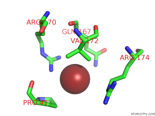
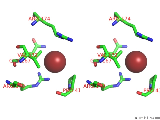
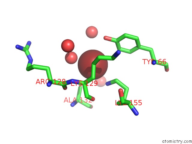
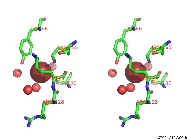
Bromine binding site 1 out of 6 in 6iyx
Go back to
Bromine binding site 1 out
of 6 in the Crystal Structure Analysis of A Eukaryotic Membrane Protein
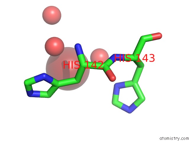
Mono view
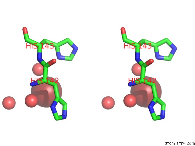
Stereo pair view
Mono view
Stereo pair view
A full contact list of Bromine with other atoms in the Br binding
site number 1 of Crystal Structure Analysis of A Eukaryotic Membrane Protein within 5.0Å range:
|
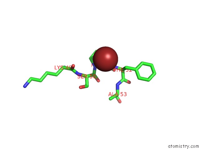
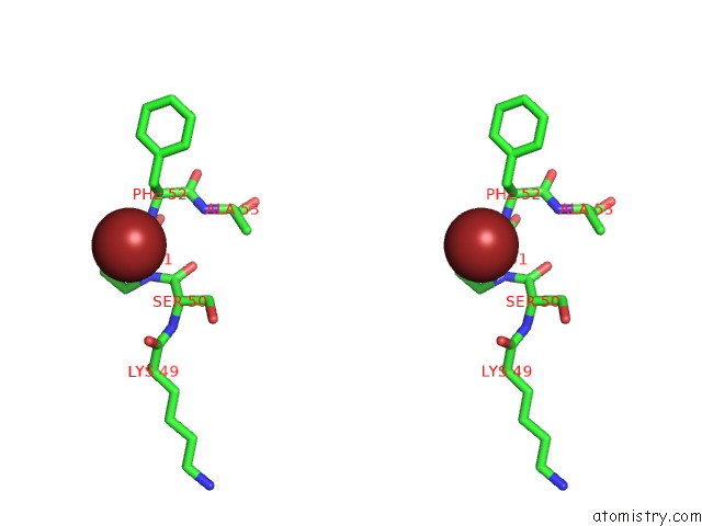
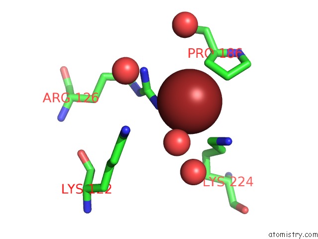
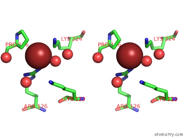
Bromine binding site 2 out of 6 in 6iyx
Go back to
Bromine binding site 2 out
of 6 in the Crystal Structure Analysis of A Eukaryotic Membrane Protein
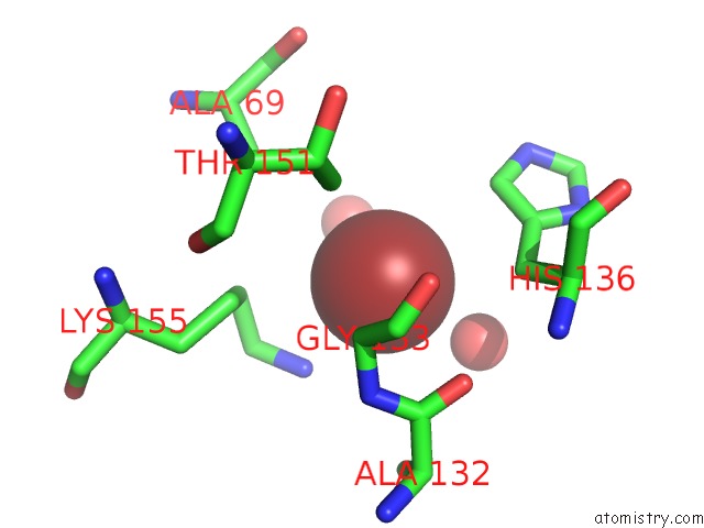
Mono view
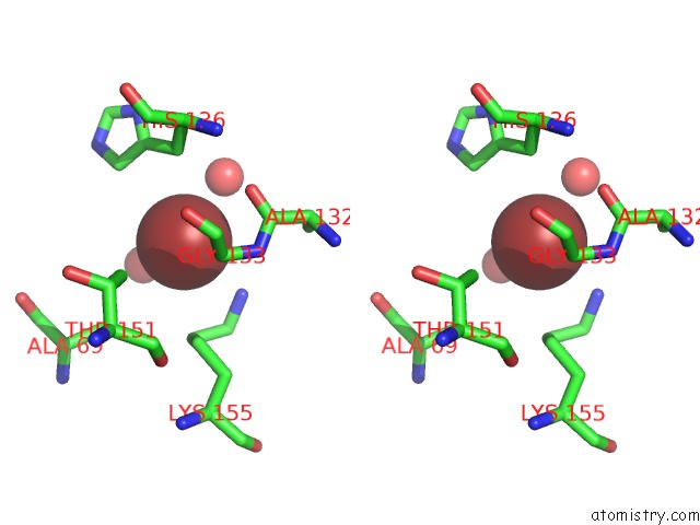
Stereo pair view
Mono view
Stereo pair view
A full contact list of Bromine with other atoms in the Br binding
site number 2 of Crystal Structure Analysis of A Eukaryotic Membrane Protein within 5.0Å range:
|
Bromine binding site 3 out of 6 in 6iyx
Go back to
Bromine binding site 3 out
of 6 in the Crystal Structure Analysis of A Eukaryotic Membrane Protein
Mono view
Stereo pair view
Mono view
Stereo pair view
A full contact list of Bromine with other atoms in the Br binding
site number 3 of Crystal Structure Analysis of A Eukaryotic Membrane Protein within 5.0Å range:
|
Bromine binding site 4 out of 6 in 6iyx
Go back to
Bromine binding site 4 out
of 6 in the Crystal Structure Analysis of A Eukaryotic Membrane Protein
Mono view
Stereo pair view
Mono view
Stereo pair view
A full contact list of Bromine with other atoms in the Br binding
site number 4 of Crystal Structure Analysis of A Eukaryotic Membrane Protein within 5.0Å range:
|
Bromine binding site 5 out of 6 in 6iyx
Go back to
Bromine binding site 5 out
of 6 in the Crystal Structure Analysis of A Eukaryotic Membrane Protein
Mono view
Stereo pair view
Mono view
Stereo pair view
A full contact list of Bromine with other atoms in the Br binding
site number 5 of Crystal Structure Analysis of A Eukaryotic Membrane Protein within 5.0Å range:
|
Bromine binding site 6 out of 6 in 6iyx
Go back to
Bromine binding site 6 out
of 6 in the Crystal Structure Analysis of A Eukaryotic Membrane Protein
Mono view
Stereo pair view
Mono view
Stereo pair view
A full contact list of Bromine with other atoms in the Br binding
site number 6 of Crystal Structure Analysis of A Eukaryotic Membrane Protein within 5.0Å range:
|
Reference:
X.H.Wang,
M.Su,
F.Gao,
W.Xie,
Y.Zeng,
D.L.Li,
X.L.Liu,
H.Zhao,
L.Qin,
F.Li,
Q.Liu,
O.B.Clarke,
S.M.Lam,
G.H.Shui,
W.A.Hendrickson,
Y.H.Chen.
Structural Basis For Activity of Tric Counter-Ion Channels in Calcium Release. Proc.Natl.Acad.Sci.Usa V. 116 4238 2019.
ISSN: ESSN 1091-6490
PubMed: 30770441
DOI: 10.1073/PNAS.1817271116
Page generated: Mon Jul 7 10:03:42 2025
ISSN: ESSN 1091-6490
PubMed: 30770441
DOI: 10.1073/PNAS.1817271116
Last articles
K in 8CTZK in 8CTX
K in 8CGV
K in 8CTS
K in 8CTT
K in 8CTN
K in 8CGK
K in 8CSU
K in 8CST
K in 8CSS