Bromine »
PDB 7cud-7fl5 »
7euw »
Bromine in PDB 7euw: X-Ray Structure of High-Strength Hydrogel-Grown FABP3 Crystal Soaked in 50% Dmso Solution Containing 4-[2-[1-(4-Bromophenyl)-5-Phenyl-1H- Pyrazol-3-Yl]Phenoxy] (HA174)
Protein crystallography data
The structure of X-Ray Structure of High-Strength Hydrogel-Grown FABP3 Crystal Soaked in 50% Dmso Solution Containing 4-[2-[1-(4-Bromophenyl)-5-Phenyl-1H- Pyrazol-3-Yl]Phenoxy] (HA174), PDB code: 7euw
was solved by
S.Sugiyama,
K.Kakinouchi,
R.Nakano,
S.Matsuoka,
H.Tsuchikawa,
M.Sonoyama,
Y.Inoue,
F.Hayashi,
M.Murata,
with X-Ray Crystallography technique. A brief refinement statistics is given in the table below:
| Resolution Low / High (Å) | 31.00 / 1.55 |
| Space group | P 21 21 21 |
| Cell size a, b, c (Å), α, β, γ (°) | 54.553, 70.421, 34.502, 90, 90, 90 |
| R / Rfree (%) | 16.2 / 20.5 |
Bromine Binding Sites:
The binding sites of Bromine atom in the X-Ray Structure of High-Strength Hydrogel-Grown FABP3 Crystal Soaked in 50% Dmso Solution Containing 4-[2-[1-(4-Bromophenyl)-5-Phenyl-1H- Pyrazol-3-Yl]Phenoxy] (HA174)
(pdb code 7euw). This binding sites where shown within
5.0 Angstroms radius around Bromine atom.
In total only one binding site of Bromine was determined in the X-Ray Structure of High-Strength Hydrogel-Grown FABP3 Crystal Soaked in 50% Dmso Solution Containing 4-[2-[1-(4-Bromophenyl)-5-Phenyl-1H- Pyrazol-3-Yl]Phenoxy] (HA174), PDB code: 7euw:
In total only one binding site of Bromine was determined in the X-Ray Structure of High-Strength Hydrogel-Grown FABP3 Crystal Soaked in 50% Dmso Solution Containing 4-[2-[1-(4-Bromophenyl)-5-Phenyl-1H- Pyrazol-3-Yl]Phenoxy] (HA174), PDB code: 7euw:
Bromine binding site 1 out of 1 in 7euw
Go back to
Bromine binding site 1 out
of 1 in the X-Ray Structure of High-Strength Hydrogel-Grown FABP3 Crystal Soaked in 50% Dmso Solution Containing 4-[2-[1-(4-Bromophenyl)-5-Phenyl-1H- Pyrazol-3-Yl]Phenoxy] (HA174)
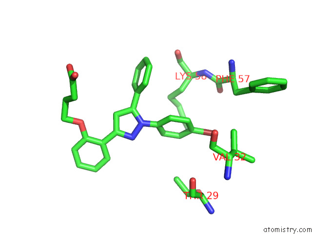
Mono view
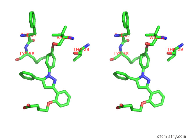
Stereo pair view
Mono view
Stereo pair view
A full contact list of Bromine with other atoms in the Br binding
site number 1 of X-Ray Structure of High-Strength Hydrogel-Grown FABP3 Crystal Soaked in 50% Dmso Solution Containing 4-[2-[1-(4-Bromophenyl)-5-Phenyl-1H- Pyrazol-3-Yl]Phenoxy] (HA174) within 5.0Å range:
|
Reference:
S.Sugiyama,
K.Kakinouchi,
S.Matsuoka,
H.Tsuchikawa,
M.Sonoyama,
Y.Inoue,
F.Hayashi,
M.Murata.
X-Ray Structure of the Human Heart Fatty Acid-Binding Protein Complexed with 4-[2-[1-(4-Bromophenyl)-5-Phenyl-1H-Pyrazol-3-Yl]Phenoxy] (HA176) To Be Published.
Page generated: Mon Jul 7 11:13:36 2025
Last articles
Na in 2WNNNa in 2WL4
Na in 2WMZ
Na in 2WLU
Na in 2WKV
Na in 2WL5
Na in 2WIS
Na in 2WKT
Na in 2WGG
Na in 2WIQ